• 文献检索
  • 文档翻译
  • 深度研究
  • 学术资讯
  • Suppr Zotero 插件Zotero 插件
  • 邀请有礼
  • 套餐&价格
  • 历史记录
应用&插件
Suppr Zotero 插件Zotero 插件浏览器插件Mac 客户端Windows 客户端微信小程序
定价
高级版会员购买积分包购买API积分包
服务
文献检索文档翻译深度研究API 文档MCP 服务
关于我们
关于 Suppr公司介绍联系我们用户协议隐私条款
关注我们

Suppr 超能文献

核心技术专利:CN118964589B侵权必究
粤ICP备2023148730 号-1Suppr @ 2026

文献检索

告别复杂PubMed语法,用中文像聊天一样搜索,搜遍4000万医学文献。AI智能推荐,让科研检索更轻松。

立即免费搜索

文件翻译

保留排版,准确专业,支持PDF/Word/PPT等文件格式,支持 12+语言互译。

免费翻译文档

深度研究

AI帮你快速写综述,25分钟生成高质量综述,智能提取关键信息,辅助科研写作。

立即免费体验

用于定量视网膜光学相干断层扫描血管造影的开源图像分析工具箱。

An Open-source Image Analysis Toolbox for Quantitative Retinal Optical Coherence Tomography Angiography.

作者信息

Amirmoezzi Yalda, Ghofrani-Jahromi Mohsen, Parsaei Hossein, Afarid Mehrdad, Mohsenipoor Negar

机构信息

Department of Medical Physics and Engineering, School of Medicine, Shiraz University of Medical Sciences, Shiraz, Iran.

Shiraz Neuroscience Research Center, Shiraz University of Medical Sciences, Shiraz, Iran.

出版信息

J Biomed Phys Eng. 2024 Feb 1;14(1):31-42. doi: 10.31661/jbpe.v0i0.2106-1349. eCollection 2024 Feb.

DOI:10.31661/jbpe.v0i0.2106-1349
PMID:38357600
原文链接:https://pmc.ncbi.nlm.nih.gov/articles/PMC10862112/
Abstract

BACKGROUND

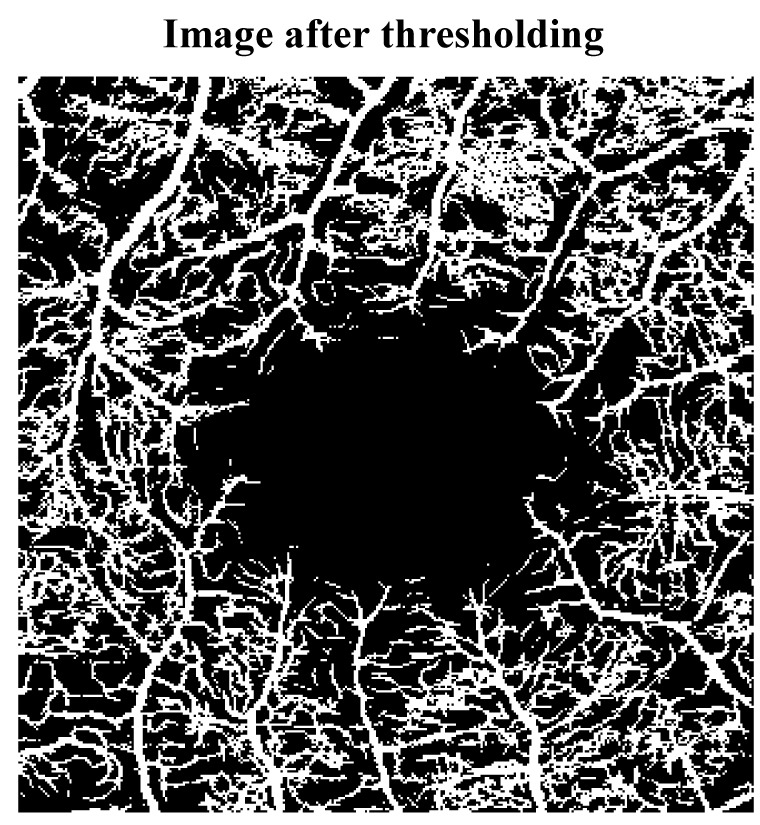
Qualitative and quantitative assessment of retinal perfusion using optical coherence tomography angiography (OCTA) has shown to be effective in the treatment and management of various retinal and optic nerve diseases. However, manual analyses of OCTA images to calculate metrics related to Foveal Avascular Zone (FAZ) morphology, and retinal vascular density and morphology are costly, time-consuming, subject to human error, and are exposed to both inter and intra operator variability.

OBJECTIVE

This study aimed to develop an open-source software framework for quantitative OCTA (QOCTA). Particularly, for analyzing OCTA images and measuring several indices describing microvascular morphology, vessel morphology, and FAZ morphology.

MATERIAL AND METHODS

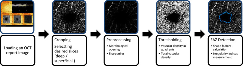
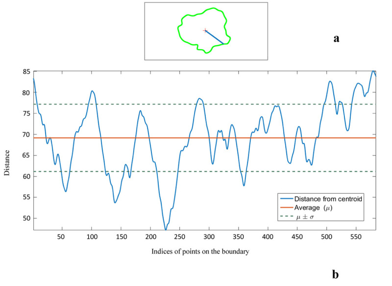
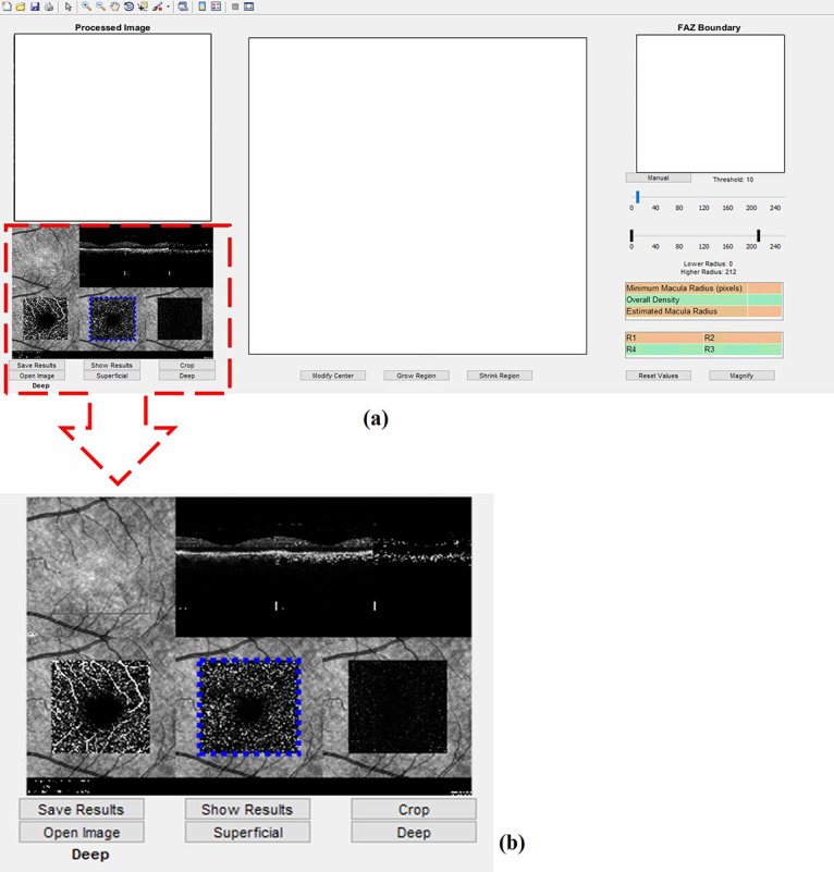
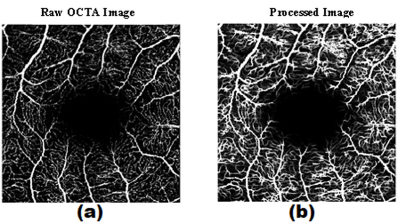
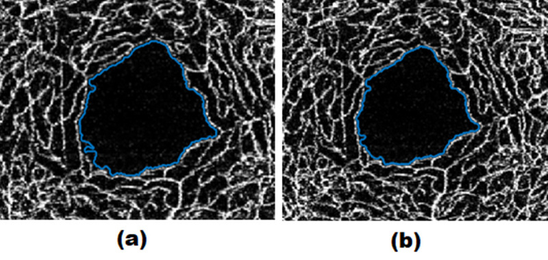
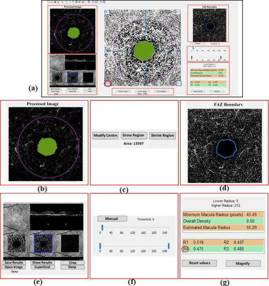
In this analytical study, we developed a toolbox or QOCTA using image processing algorithms provided in MATLAB. The software automatically determines FAZ and measures several parameters related to both size and shape of FAZ including area, perimeter, Feret's diameter circularity, axial ratio, roundness, and solidity. The microvascular structure is derived from the processed image to estimate the vessel density (VD). To assess the reliability of the software, three independent operators measured the mentioned parameters for the eyes of 21 subjects. The consistency of the values was assessed using the intraclass correlation coefficient (ICC) index.

RESULTS

Excellent consistency was observed between the measurements completed for the superficial layer, ICC >0.9. For the deep layer, good reliability in the measurements was achieved, ICC >0.7.

CONCLUSION

The developed software is reliable; hence, it can facilitate quantitative OCTA, further statistical comparison in cohort OCTA studies, and can assist with obtaining deeper insights into retinal variations in various populations.

摘要

背景

使用光学相干断层扫描血管造影(OCTA)对视网膜灌注进行定性和定量评估已被证明在各种视网膜和视神经疾病的治疗与管理中是有效的。然而,通过手动分析OCTA图像来计算与黄斑无血管区(FAZ)形态、视网膜血管密度和形态相关的指标,成本高、耗时、易出现人为误差,并且存在操作者间和操作者内的变异性。

目的

本研究旨在开发一个用于定量OCTA(QOCTA)的开源软件框架。特别是用于分析OCTA图像并测量描述微血管形态、血管形态和FAZ形态的几个指标。

材料与方法

在这项分析研究中,我们使用MATLAB中提供的图像处理算法开发了一个QOCTA工具箱。该软件自动确定FAZ并测量与FAZ的大小和形状相关的几个参数,包括面积、周长、费雷特直径、圆形度、轴比、圆度和紧实度。从处理后的图像中得出微血管结构以估计血管密度(VD)。为了评估该软件的可靠性,三名独立的操作者对21名受试者的眼睛测量了上述参数。使用组内相关系数(ICC)指数评估值的一致性。

结果

在浅层测量之间观察到极好的一致性,ICC>0.9。对于深层,测量具有良好的可靠性,ICC>0.7。

结论

所开发的软件是可靠的;因此,它可以促进定量OCTA、队列OCTA研究中的进一步统计比较,并有助于更深入地了解不同人群的视网膜变化。

https://cdn.ncbi.nlm.nih.gov/pmc/blobs/c645/10862112/9c597237ee3b/JBPE-14-31-g007.jpg
https://cdn.ncbi.nlm.nih.gov/pmc/blobs/c645/10862112/bb6ace059e22/JBPE-14-31-g001.jpg
https://cdn.ncbi.nlm.nih.gov/pmc/blobs/c645/10862112/64a29c658a74/JBPE-14-31-g002.jpg
https://cdn.ncbi.nlm.nih.gov/pmc/blobs/c645/10862112/20479e79ae20/JBPE-14-31-g003.jpg
https://cdn.ncbi.nlm.nih.gov/pmc/blobs/c645/10862112/7d40e7795452/JBPE-14-31-g004.jpg
https://cdn.ncbi.nlm.nih.gov/pmc/blobs/c645/10862112/3a16c00a0f41/JBPE-14-31-g005.jpg
https://cdn.ncbi.nlm.nih.gov/pmc/blobs/c645/10862112/3554ab8f9bc1/JBPE-14-31-g006.jpg
https://cdn.ncbi.nlm.nih.gov/pmc/blobs/c645/10862112/9c597237ee3b/JBPE-14-31-g007.jpg

相似文献

1
An Open-source Image Analysis Toolbox for Quantitative Retinal Optical Coherence Tomography Angiography.用于定量视网膜光学相干断层扫描血管造影的开源图像分析工具箱。
J Biomed Phys Eng. 2024 Feb 1;14(1):31-42. doi: 10.31661/jbpe.v0i0.2106-1349. eCollection 2024 Feb.
2
Evaluating the Quantitative Foveal Avascular Zone and Retino-Choroidal Vessel Density Using Optical Coherence Tomography Angiography in a Healthy Indian Population.在健康印度人群中使用光学相干断层扫描血管造影术评估定量黄斑无血管区和视网膜脉络膜血管密度
Cureus. 2022 Aug 4;14(8):e27669. doi: 10.7759/cureus.27669. eCollection 2022 Aug.
3
Factors Affecting Repeatability of Foveal Avascular Zone Measurement Using Optical Coherence Tomography Angiography in Pathologic Eyes.影响光学相干断层扫描血管造影术测量病理性眼黄斑无血管区重复性的因素
Clin Ophthalmol. 2020 Apr 5;14:1025-1033. doi: 10.2147/OPTH.S247172. eCollection 2020.
4
Repeatability, interocular correlation and agreement of quantitative swept-source optical coherence tomography angiography macular metrics in healthy subjects.健康受试者定量扫频源光学相干断层血管造影黄斑参数的可重复性、眼间相关性和一致性。
Br J Ophthalmol. 2019 Mar;103(3):415-420. doi: 10.1136/bjophthalmol-2018-311874. Epub 2018 May 29.
5
Repeatability of Wide-field Optical Coherence Tomography Angiography in Normal Retina.正常视网膜广角光学相干断层扫描血管造影的可重复性
Transl Vis Sci Technol. 2019 May 2;8(3):6. doi: 10.1167/tvst.8.3.6. eCollection 2019 May.
6
Improved Automated Foveal Avascular Zone Measurement in Cirrus Optical Coherence Tomography Angiography Using the Level Sets Macro.利用水平集宏在 Cirrus 光学相干断层扫描血管造影中进行改进的自动黄斑无血管区测量。
Transl Vis Sci Technol. 2020 Nov 13;9(12):20. doi: 10.1167/tvst.9.12.20. eCollection 2020 Nov.
7
A standardized method to quantitatively analyze optical coherence tomography angiography images of the macular and peripapillary vessels.一种定量分析黄斑和视乳头周围血管光学相干断层扫描血管造影图像的标准化方法。
Int J Retina Vitreous. 2022 Oct 15;8(1):75. doi: 10.1186/s40942-022-00426-9.
8
Macular vessels density in diabetic retinopathy: quantitative assessment using optical coherence tomography angiography.糖尿病视网膜病变中的黄斑血管密度:使用光学相干断层扫描血管造影术进行定量评估。
Int Ophthalmol. 2019 Aug;39(8):1845-1859. doi: 10.1007/s10792-018-1013-0. Epub 2018 Sep 7.
9
Reproducibility of Foveal Avascular Zone and Superficial Macular Retinal Vasculature Measurements in Healthy Eyes Determined by Two Different Scanning Protocols of Optical Coherence Tomography Angiography.两种不同的光学相干断层扫描血管造影扫描方案在健康眼中对中心凹无血管区和浅层视网膜脉络膜血管测量的重复性。
Ophthalmic Res. 2020;63(3):244-251. doi: 10.1159/000503071. Epub 2019 Oct 16.
10
Evaluation of microvascular network with optical coherence tomography angiography (OCTA) in branch retinal vein occlusion (BRVO).应用光相干断层扫描血管造影术(OCTA)评估分支视网膜静脉阻塞(BRVO)的微血管网络。
BMC Ophthalmol. 2020 Apr 19;20(1):154. doi: 10.1186/s12886-020-01405-0.

引用本文的文献

1
DERMA-OCTA: A Comprehensive Dataset and Preprocessing Pipeline for Dermatological OCTA Vessel Segmentation.DERMA-OCTA:用于皮肤病光学相干断层扫描血管造影(OCTA)血管分割的综合数据集及预处理流程
Sci Data. 2025 Aug 23;12(1):1473. doi: 10.1038/s41597-025-05763-6.
2
Low rank based FAZ segmentation in OCTA images.光学相干断层扫描血管造影(OCTA)图像中基于低秩的黄斑无血管区(FAZ)分割
Sci Rep. 2025 Jul 9;15(1):24757. doi: 10.1038/s41598-025-07448-x.
3
OCTA-ReVA: an open-source toolbox for comprehensive retinal vessel feature analysis in optical coherence tomography angiography.

本文引用的文献

1
Quantification of retinal microvascular parameters by severity of diabetic retinopathy using wide-field swept-source optical coherence tomography angiography.采用广角扫频源光学相干断层血管造影术根据糖尿病视网膜病变严重程度定量分析视网膜微血管参数。
Graefes Arch Clin Exp Ophthalmol. 2021 Aug;259(8):2103-2111. doi: 10.1007/s00417-021-05099-y. Epub 2021 Feb 2.
2
Quantitative analysis of retinal and choriocapillary vascular density of multiple evanescent white dot syndrome by optical coherence tomography angiography.光学相干断层扫描血管造影定量分析多发性一过性白点综合征的视网膜及脉络膜毛细血管密度。
Graefes Arch Clin Exp Ophthalmol. 2020 Aug;258(8):1697-1707. doi: 10.1007/s00417-020-04687-8. Epub 2020 Apr 29.
3
OCTA-ReVA:一种用于光学相干断层扫描血管造影中视网膜血管特征综合分析的开源工具箱。
Biomed Opt Express. 2024 Sep 25;15(10):6010-6023. doi: 10.1364/BOE.537727. eCollection 2024 Oct 1.
4
Towards standardising retinal OCT angiography image analysis with open-source toolbox OCTAVA.用开源工具 OCTAVA 实现视网膜 OCT 血管造影图像分析标准化。
Sci Rep. 2024 Mar 12;14(1):5979. doi: 10.1038/s41598-024-53501-6.
5
Assessment of macular findings by OCT angiography in patients without clinical signs of diabetic retinopathy: radiomics features for early screening of diabetic retinopathy.OCT 血管造影评估无临床糖尿病视网膜病变征象患者的黄斑病变:用于糖尿病视网膜病变早期筛查的放射组学特征。
BMC Ophthalmol. 2022 Jun 27;22(1):281. doi: 10.1186/s12886-022-02492-x.
Objective evaluation of size and shape of superficial foveal avascular zone in normal subjects by optical coherence tomography angiography.
应用光相干断层扫描血管造影术客观评价正常人群浅层中心凹无血管区的大小和形态。
Sci Rep. 2018 Jul 4;8(1):10143. doi: 10.1038/s41598-018-28530-7.
4
Optical coherence tomography angiography: Technical principles and clinical applications in ophthalmology.光学相干断层扫描血管造影:眼科的技术原理与临床应用
Taiwan J Ophthalmol. 2017 Jul-Sep;7(3):115-129. doi: 10.4103/tjo.tjo_31_17. Epub 2017 Sep 19.
5
Method for quantitative assessment of retinal vessel tortuosity in optical coherence tomography angiography applied to sickle cell retinopathy.应用于镰状细胞视网膜病变的光学相干断层扫描血管造影术中视网膜血管迂曲度的定量评估方法
Biomed Opt Express. 2017 Jul 24;8(8):3796-3806. doi: 10.1364/BOE.8.003796. eCollection 2017 Aug 1.
6
Determinants of Quantitative Optical Coherence Tomography Angiography Metrics in Patients with Diabetes.糖尿病患者定量光学相干断层扫描血管造影测量指标的影响因素。
Sci Rep. 2017 May 31;7(1):2575. doi: 10.1038/s41598-017-02767-0.
7
A review of optical coherence tomography angiography (OCTA).光学相干断层扫描血管造影术(OCTA)综述。
Int J Retina Vitreous. 2015 Apr 15;1:5. doi: 10.1186/s40942-015-0005-8. eCollection 2015.
8
Retinal Capillary Network and Foveal Avascular Zone in Eyes with Vein Occlusion and Fellow Eyes Analyzed With Optical Coherence Tomography Angiography.光学相干断层扫描血管造影分析静脉阻塞患眼及对侧眼的视网膜毛细血管网络和黄斑无血管区
Invest Ophthalmol Vis Sci. 2016 Jul 1;57(9):OCT486-94. doi: 10.1167/iovs.15-18907.
9
Quantifying Microvascular Density and Morphology in Diabetic Retinopathy Using Spectral-Domain Optical Coherence Tomography Angiography.使用频域光学相干断层扫描血管造影术量化糖尿病视网膜病变中的微血管密度和形态。
Invest Ophthalmol Vis Sci. 2016 Jul 1;57(9):OCT362-70. doi: 10.1167/iovs.15-18904.
10
A Guideline of Selecting and Reporting Intraclass Correlation Coefficients for Reliability Research.可靠性研究中组内相关系数选择与报告指南
J Chiropr Med. 2016 Jun;15(2):155-63. doi: 10.1016/j.jcm.2016.02.012. Epub 2016 Mar 31.