Department of Gastrointestinal Surgery, The Second Hospital of Anhui Medical University, Hefei, 230601, China.
Bariatric Center, the Second Hospital of Anhui Medical University, Hefei, 230601, China.
J Neuroinflammation. 2022 Jun 27;19(1):166. doi: 10.1186/s12974-022-02529-4.
Little is known about how the obesogenic environment influences emotional states associated with glial responses and neuronal function. Here, we investigated glial reactivation and neuronal electrophysiological properties in emotion-related brain regions of high-fat diet (HFD) and ob/ob mice under chronic stress.
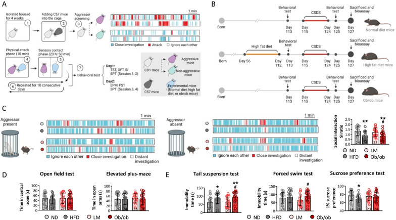
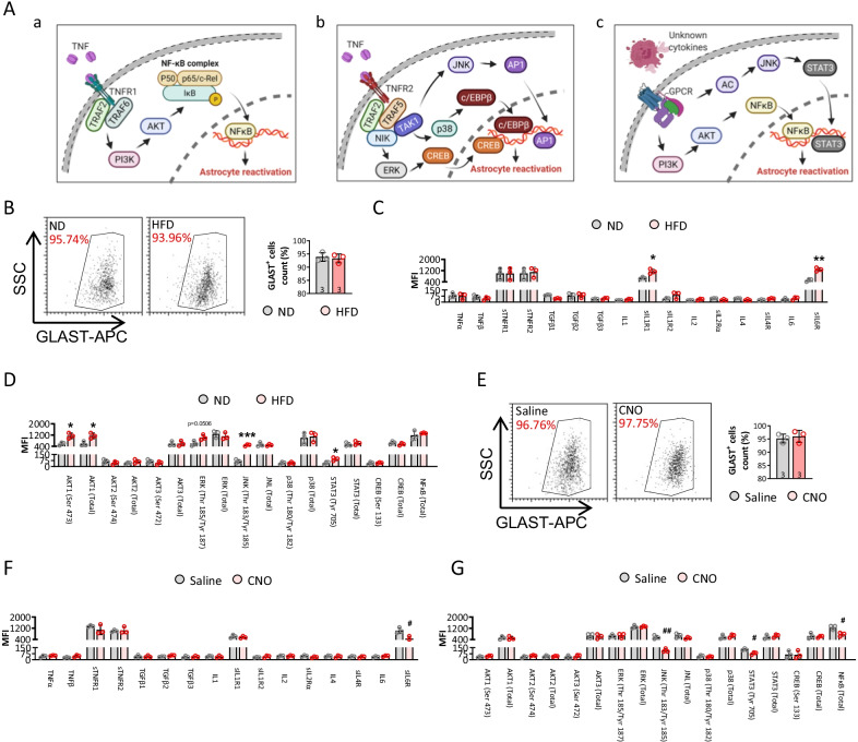
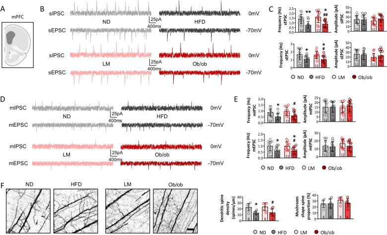
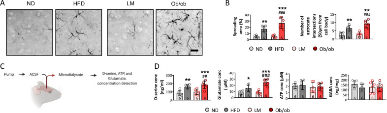
The glial reactivation and neuronal activities in emotion-related brain regions were analyzed among normal diet mice (ND), HFD mice, wild-type mice, and ob/ob mice. To further activate or inhibit astrocytes in medial prefrontal cortex (mPFC), we injected astrocytes specific Gq-AAV or Gi-AAV into mPFC and ongoing treated mice with CNO.
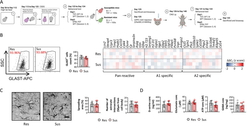
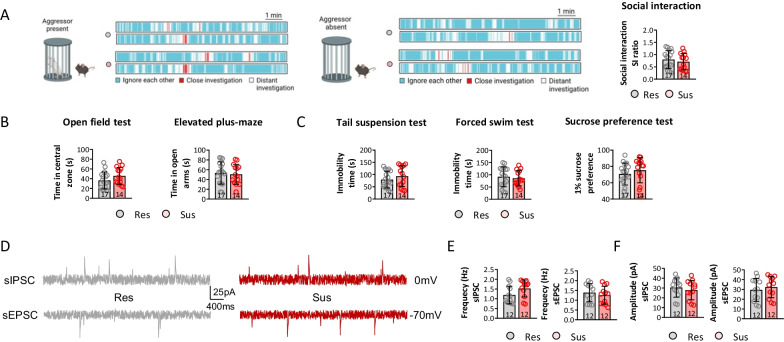
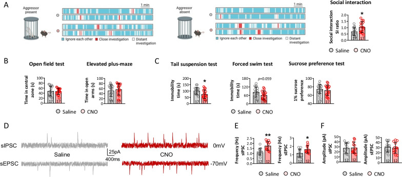
The results showed that obesogenic factors per se had no significant effect on neuronal activities in emotion-related brain regions, or on behavioral performance. However, exposure to a chronic stressor profoundly reduced the frequency of spontaneous inhibitory postsynaptic currents (sIPSCs) and spontaneous excitatory postsynaptic currents (sEPSCs) in the mPFC; depressive-like behaviors were seen, accompanied by significant upregulation of astrocyte reactivation. We identified resilient and susceptible mice among chronic social defeat stress-exposed HFD mice. As expected, astrocyte reactivity was upregulated, while neuronal activity was depressed, in the mPFC of susceptible compared to resilient mice. Furthermore, activating astrocytes resulted in similar levels of neuronal activity and depressive-like behaviors between resilient and susceptible mice. Additionally, inhibiting astrocyte reactivation in the mPFC of HFD mice upregulated neuronal activities and inhibited depressive-like behaviors.
These observations indicate that obesogenic factors increase the risk of depression, and improve our understanding of the pathological relationship between obesity and depression.
肥胖环境如何影响与神经胶质反应和神经元功能相关的情绪状态知之甚少。在这里,我们研究了高脂肪饮食(HFD)和 ob/ob 小鼠在慢性应激下与情绪相关的大脑区域中神经胶质的再激活和神经元电生理特性。
在正常饮食小鼠(ND)、HFD 小鼠、野生型小鼠和 ob/ob 小鼠中分析了与情绪相关的大脑区域中的神经胶质再激活和神经元活动。为了进一步激活或抑制前额叶皮质(mPFC)中的星形胶质细胞,我们将特异性 Gq-AAV 或 Gi-AAV 注入 mPFC 并持续用 CNO 处理小鼠。
结果表明,肥胖因素本身对与情绪相关的大脑区域中的神经元活动或行为表现没有显著影响。然而,暴露于慢性应激源会严重降低 mPFC 中自发性抑制性突触后电流(sIPSCs)和自发性兴奋性突触后电流(sEPSCs)的频率;出现抑郁样行为,同时星形胶质细胞再激活显著上调。我们在慢性社交挫败应激暴露的 HFD 小鼠中鉴定出有弹性和易感的小鼠。正如预期的那样,与有弹性的小鼠相比,易感的小鼠 mPFC 中的星形胶质细胞反应性上调,而神经元活性被抑制。此外,激活星形胶质细胞会导致有弹性和易感的小鼠之间具有相似水平的神经元活性和抑郁样行为。此外,抑制 HFD 小鼠 mPFC 中的星形胶质细胞再激活会增加神经元活性并抑制抑郁样行为。
这些观察结果表明肥胖因素会增加患抑郁症的风险,并增进我们对肥胖和抑郁症之间病理关系的理解。