Division of Pediatric Rheumatology, Department of Pediatrics, University of Michigan, Ann Arbor.
Department of Chemical Engineering, University of Michigan, Ann Arbor.
Arthritis Rheumatol. 2022 Dec;74(12):2024-2031. doi: 10.1002/art.42283. Epub 2022 Oct 18.
Cutaneous inflammation can signal disease in juvenile dermatomyositis (DM) and childhood-onset systemic lupus erythematosus (cSLE), but we do not fully understand cellular mechanisms of cutaneous inflammation. In this study, we used imaging mass cytometry to characterize cutaneous inflammatory cell populations and cell-cell interactions in juvenile DM as compared to cSLE.
We performed imaging mass cytometry analysis on skin biopsy samples from juvenile DM patients (n = 6) and cSLE patients (n = 4). Tissue slides were processed and incubated with metal-tagged antibodies for CD14, CD15, CD16, CD56, CD68, CD11c, HLA-DR, blood dendritic cell antigen 2, CD20, CD27, CD138, CD4, CD8, E-cadherin, CD31, pan-keratin, and type I collagen. Stained tissue was ablated, and raw data were acquired using the Hyperion imaging system. We utilized the Phenograph unsupervised clustering algorithm to determine cell marker expression and permutation test by histoCAT to perform neighborhood analysis.
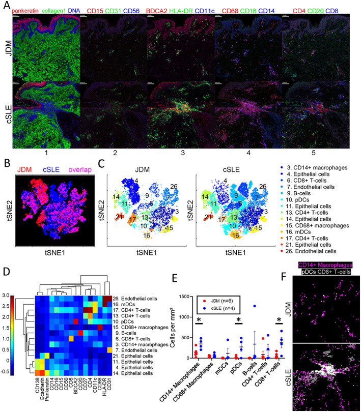
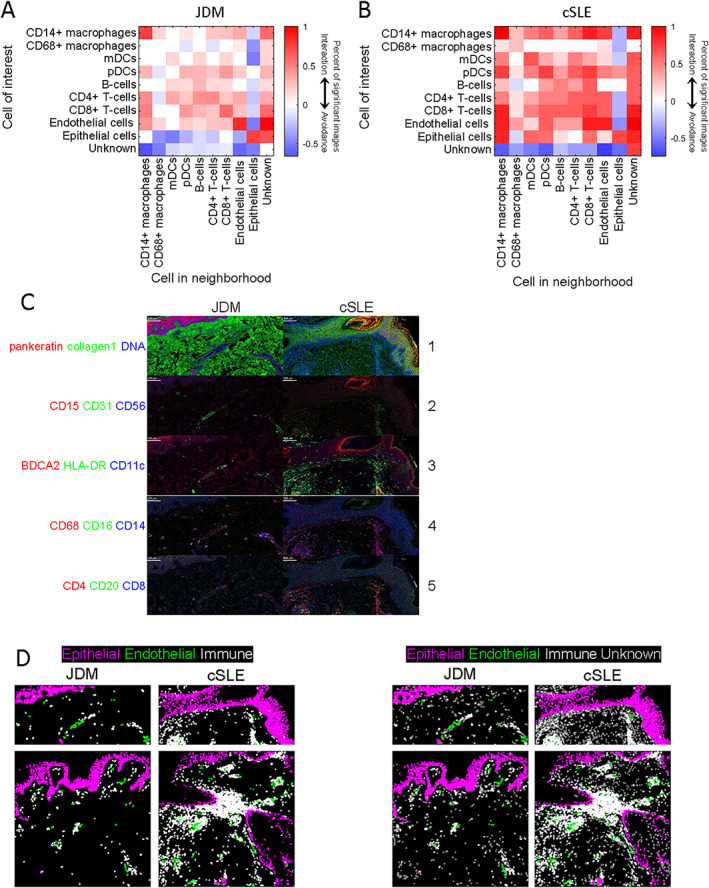
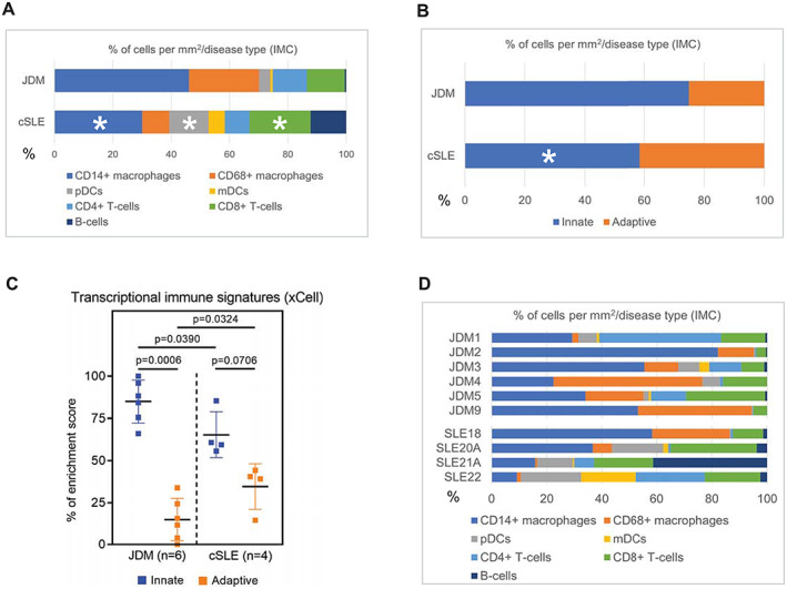
We identified 14 cell populations in juvenile DM and cSLE skin, including CD14+ and CD68+ macrophages, myeloid and plasmacytoid dendritic cells (pDCs), CD4+ and CD8+ T cells, and B cells. Overall, cSLE skin had a higher inflammatory cell infiltrate, with increased CD14+ macrophages, pDCs, and CD8+ T cells and immune cell-immune cell interactions. Juvenile DM skin displayed a stronger innate immune signature, with a higher overall percentage of CD14+ macrophages and prominent endothelial cell-immune cell interaction.
Our findings identify immune cell population differences, including CD14+ macrophages, pDCs, and CD8+ T cells, in juvenile DM skin compared to cSLE skin, and highlight a predominant innate immune signature and endothelial cell-immune cell interaction in juvenile DM, providing insight into candidate cell populations and interactions to better understand disease-specific pathophysiology.
皮肤炎症可提示幼年皮肌炎(juvenile dermatomyositis,DM)和儿童发病的系统性红斑狼疮(childhood-onset systemic lupus erythematosus,cSLE)的疾病情况,但我们尚未完全了解皮肤炎症的细胞机制。在本研究中,我们通过成像质谱细胞术(imaging mass cytometry)来比较幼年 DM 和 cSLE 患者的皮肤炎症细胞群和细胞间相互作用。
我们对 6 例幼年 DM 患者和 4 例 cSLE 患者的皮肤活检样本进行了成像质谱细胞术分析。组织切片经过处理并用金属标记的抗体孵育,这些抗体包括 CD14、CD15、CD16、CD56、CD68、CD11c、HLA-DR、血液树突状细胞抗原 2、CD20、CD27、CD138、CD4、CD8、E-钙黏蛋白、CD31、广谱角蛋白和 I 型胶原。染色组织被消融,并用 Hyperion 成像系统采集原始数据。我们利用 Phenograph 无监督聚类算法确定细胞标志物的表达,并通过 histoCAT 进行排列检验以执行邻域分析。
我们在幼年 DM 和 cSLE 皮肤中鉴定出 14 种细胞群,包括 CD14+和 CD68+巨噬细胞、髓样和浆细胞样树突状细胞(plasmacytoid dendritic cells,pDCs)、CD4+和 CD8+T 细胞以及 B 细胞。总的来说,cSLE 皮肤有更高的炎症细胞浸润,表现为 CD14+巨噬细胞、pDCs 和 CD8+T 细胞以及免疫细胞-免疫细胞相互作用增加。幼年 DM 皮肤表现出更强的固有免疫特征,CD14+巨噬细胞的总体百分比更高,并且突出表现为内皮细胞-免疫细胞相互作用。
与 cSLE 皮肤相比,我们的研究结果在幼年 DM 皮肤中发现了免疫细胞群体的差异,包括 CD14+巨噬细胞、pDCs 和 CD8+T 细胞,并强调了幼年 DM 中主要的固有免疫特征和内皮细胞-免疫细胞相互作用,为更好地了解疾病特异性病理生理学提供了候选细胞群和相互作用的见解。