• 文献检索
  • 文档翻译
  • 深度研究
  • 学术资讯
  • Suppr Zotero 插件Zotero 插件
  • 邀请有礼
  • 套餐&价格
  • 历史记录
应用&插件
Suppr Zotero 插件Zotero 插件浏览器插件Mac 客户端Windows 客户端微信小程序
定价
高级版会员购买积分包购买API积分包
服务
文献检索文档翻译深度研究API 文档MCP 服务
关于我们
关于 Suppr公司介绍联系我们用户协议隐私条款
关注我们

Suppr 超能文献

核心技术专利:CN118964589B侵权必究
粤ICP备2023148730 号-1Suppr @ 2026

文献检索

告别复杂PubMed语法,用中文像聊天一样搜索,搜遍4000万医学文献。AI智能推荐,让科研检索更轻松。

立即免费搜索

文件翻译

保留排版,准确专业,支持PDF/Word/PPT等文件格式,支持 12+语言互译。

免费翻译文档

深度研究

AI帮你快速写综述,25分钟生成高质量综述,智能提取关键信息,辅助科研写作。

立即免费体验

基于成像流式细胞术的方法,用于分析未经处理的人血浆中的单个细胞外囊泡。

An imaging flow cytometry-based methodology for the analysis of single extracellular vesicles in unprocessed human plasma.

机构信息

Erasmus MC Transplant Institute, Department of Internal Medicine, University Medical Center Rotterdam, Rotterdam, The Netherlands.

Biomedical Engineering & Physics, Laboratory Experimental Clinical Chemistry, Vesicle Observation Center, Amsterdam UMC, University of Amsterdam, Amsterdam, The Netherlands.

出版信息

Commun Biol. 2022 Jun 29;5(1):633. doi: 10.1038/s42003-022-03569-5.

DOI:10.1038/s42003-022-03569-5
PMID:35768629
原文链接:https://pmc.ncbi.nlm.nih.gov/articles/PMC9243126/
Abstract

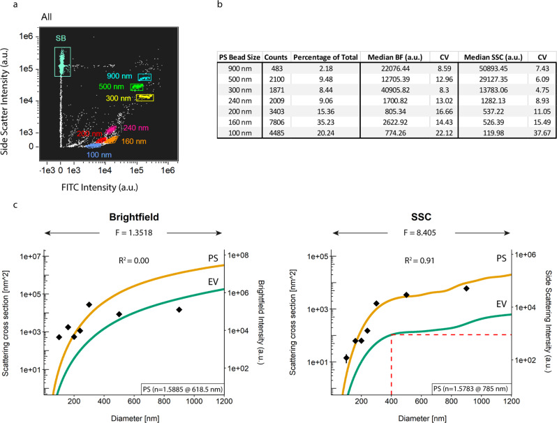
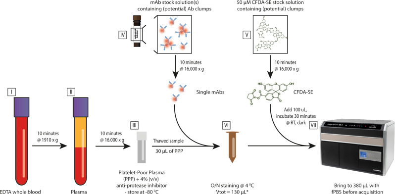
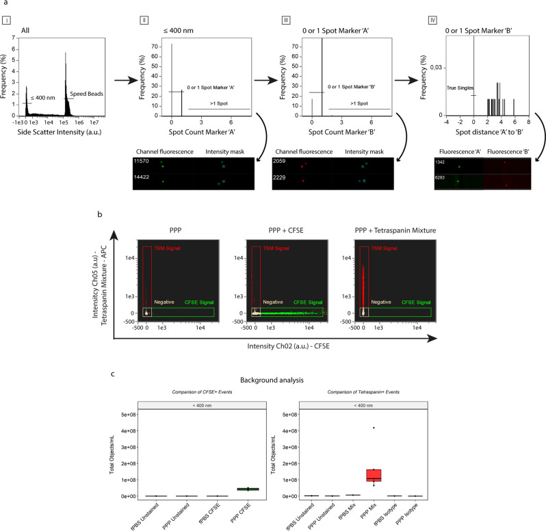
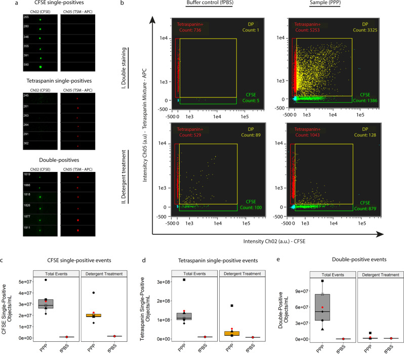
Extracellular vesicles (EVs) are tissue-specific particles released by cells containing valuable diagnostic information in the form of various biomolecules. To rule out selection bias or introduction of artefacts caused by EV isolation techniques, we present a clinically feasible, imaging flow cytometry (IFCM)-based methodology to phenotype and determine the concentration of EVs with a diameter ≤400 nm in human platelet-poor plasma (PPP) without prior isolation of EVs. Instrument calibration (both size and fluorescence) were performed with commercial polystyrene beads. Detergent treatment of EVs was performed to discriminate true vesicular events from artefacts. Using a combination of markers (CFSE & Tetraspanins, or CD9 & CD31) we found that >90% of double-positive fluorescent events represented single EVs. Through this work, we provide a framework that will allow the application of IFCM for EV analysis in peripheral blood plasma in a plethora of experimental and potentially diagnostic settings. Additionally, this direct approach for EV analysis will enable researchers to explore corners of EVs as cellular messengers in healthy and pathological conditions.

摘要

细胞外囊泡(EVs)是由细胞释放的具有组织特异性的颗粒,其中包含各种生物分子形式的有价值的诊断信息。为了排除 EV 分离技术引起的选择偏差或引入假象,我们提出了一种临床可行的、基于成像流式细胞术(IFCM)的方法,用于表型分析和确定人血小板贫乏血浆(PPP)中直径≤400nm 的 EV 的浓度,而无需事先分离 EV。仪器校准(大小和荧光)均使用商业聚苯乙烯珠进行。用去污剂处理 EVs 以区分真实的囊泡事件和假象。通过使用组合标记物(CFSE 和四跨膜蛋白,或 CD9 和 CD31),我们发现>90%的双阳性荧光事件代表单个 EVs。通过这项工作,我们提供了一个框架,允许在众多实验和潜在诊断环境中在外周血浆中应用 IFCM 进行 EV 分析。此外,这种 EV 分析的直接方法将使研究人员能够探索 EVs 在健康和病理条件下作为细胞信使的各个方面。

https://cdn.ncbi.nlm.nih.gov/pmc/blobs/d7f8/9243126/15eb12c0eccd/42003_2022_3569_Fig9_HTML.jpg
https://cdn.ncbi.nlm.nih.gov/pmc/blobs/d7f8/9243126/704dcd091c6d/42003_2022_3569_Fig1_HTML.jpg
https://cdn.ncbi.nlm.nih.gov/pmc/blobs/d7f8/9243126/e0f4ac6b3d48/42003_2022_3569_Fig2_HTML.jpg
https://cdn.ncbi.nlm.nih.gov/pmc/blobs/d7f8/9243126/2f9b92290b8e/42003_2022_3569_Fig3_HTML.jpg
https://cdn.ncbi.nlm.nih.gov/pmc/blobs/d7f8/9243126/3ce774d04d1b/42003_2022_3569_Fig4_HTML.jpg
https://cdn.ncbi.nlm.nih.gov/pmc/blobs/d7f8/9243126/d2903982cc88/42003_2022_3569_Fig5_HTML.jpg
https://cdn.ncbi.nlm.nih.gov/pmc/blobs/d7f8/9243126/ecb7b76bc5b1/42003_2022_3569_Fig6_HTML.jpg
https://cdn.ncbi.nlm.nih.gov/pmc/blobs/d7f8/9243126/ed955510cae0/42003_2022_3569_Fig7_HTML.jpg
https://cdn.ncbi.nlm.nih.gov/pmc/blobs/d7f8/9243126/a431f94d62fd/42003_2022_3569_Fig8_HTML.jpg
https://cdn.ncbi.nlm.nih.gov/pmc/blobs/d7f8/9243126/15eb12c0eccd/42003_2022_3569_Fig9_HTML.jpg

相似文献

1
An imaging flow cytometry-based methodology for the analysis of single extracellular vesicles in unprocessed human plasma.基于成像流式细胞术的方法,用于分析未经处理的人血浆中的单个细胞外囊泡。
Commun Biol. 2022 Jun 29;5(1):633. doi: 10.1038/s42003-022-03569-5.
2
Size and fluorescence calibrated imaging flow cytometry: From arbitrary to standard units.大小和荧光校准成像流式细胞术:从任意单位到标准单位。
Cytometry A. 2024 Oct;105(10):752-762. doi: 10.1002/cyto.a.24895. Epub 2024 Sep 5.
3
Large Extracellular Vesicles Can be Characterised by Multiplex Labelling Using Imaging Flow Cytometry.大型细胞外囊泡可通过成像流式细胞术进行多重标记来表征。
Int J Mol Sci. 2020 Nov 18;21(22):8723. doi: 10.3390/ijms21228723.
4
Flow cytometric analysis of extracellular vesicle subsets in plasma: impact of swarm by particles of non-interest.流式细胞术分析血浆中细胞外囊泡亚群:非感兴趣颗粒对群体的影响。
J Thromb Haemost. 2018 Jul;16(7):1423-1436. doi: 10.1111/jth.14154. Epub 2018 Jun 15.
5
Extracellular Vesicles Released During Normothermic Machine Perfusion Are Associated With Human Donor Kidney Characteristics.在常温机器灌注过程中释放的细胞外囊泡与人类供体肾脏特征相关。
Transplantation. 2022 Dec 1;106(12):2360-2369. doi: 10.1097/TP.0000000000004215. Epub 2022 Nov 22.
6
Standardization of extracellular vesicle measurements by flow cytometry through vesicle diameter approximation.通过囊泡直径逼近实现流式细胞术对外泌体测量的标准化。
J Thromb Haemost. 2018 Jun;16(6):1236-1245. doi: 10.1111/jth.14009. Epub 2018 Apr 26.
7
Tetraspanins distinguish separate extracellular vesicle subpopulations in human serum and plasma - Contributions of platelet extracellular vesicles in plasma samples.四跨膜蛋白将人血清和血浆中的不同细胞外囊泡亚群区分开来——血小板细胞外囊泡在血浆样本中的作用。
J Extracell Vesicles. 2022 May;11(5):e12213. doi: 10.1002/jev2.12213.
8
Direct detection of circulating donor-derived extracellular vesicles in kidney transplant recipients.直接检测肾移植受者循环供体来源的细胞外囊泡。
Sci Rep. 2022 Dec 20;12(1):21973. doi: 10.1038/s41598-022-26580-6.
9
Isolation-free measurement of single urinary extracellular vesicles by imaging flow cytometry.无分离式检测:应用成像流式细胞术检测单个尿细胞外囊泡。
Nanomedicine. 2023 Feb;48:102638. doi: 10.1016/j.nano.2022.102638. Epub 2022 Dec 19.
10
Optimisation of imaging flow cytometry for the analysis of single extracellular vesicles by using fluorescence-tagged vesicles as biological reference material.通过使用荧光标记囊泡作为生物参考材料优化成像流式细胞术用于分析单个细胞外囊泡。
J Extracell Vesicles. 2019 Mar 21;8(1):1587567. doi: 10.1080/20013078.2019.1587567. eCollection 2019.

引用本文的文献

1
Central Nervous System-Derived Extracellular Vesicles as Biomarkers in Alzheimer's Disease.中枢神经系统衍生的细胞外囊泡作为阿尔茨海默病的生物标志物
Int J Mol Sci. 2025 Aug 26;26(17):8272. doi: 10.3390/ijms26178272.
2
Kidney-derived Urinary Extracellular Vesicles are Increased During Acute Rejection after Kidney Transplantation: A Pilot Study.肾移植术后急性排斥反应期间肾脏来源的尿液细胞外囊泡增加:一项初步研究。
Transplant Direct. 2025 Apr 10;11(5):e1796. doi: 10.1097/TXD.0000000000001796. eCollection 2025 May.
3
A Simple and Scalable Assay for Multiplexed Flow Cytometric Profiling of Surface Markers on Small Extracellular Vesicles.

本文引用的文献

1
Comparing Approaches to Normalize, Quantify, and Characterize Urinary Extracellular Vesicles.比较标准化、量化和表征尿细胞外囊泡的方法。
J Am Soc Nephrol. 2021 May 3;32(5):1210-1226. doi: 10.1681/ASN.2020081142. Epub 2021 Mar 29.
2
Methamphetamine use alters human plasma extracellular vesicles and their microRNA cargo: An exploratory study.甲基苯丙胺使用改变了人体血浆细胞外囊泡及其 microRNA 货物:一项探索性研究。
J Extracell Vesicles. 2020 Nov;10(1):e12028. doi: 10.1002/jev2.12028. Epub 2020 Nov 28.
3
Tetraspanins, More than Markers of Extracellular Vesicles in Reproduction.
一种用于小细胞外囊泡表面标志物多重流式细胞术分析的简单且可扩展的检测方法。
Cells. 2025 Jun 28;14(13):989. doi: 10.3390/cells14130989.
4
Tumor-derived exosomes and their application in cancer treatment.肿瘤衍生的外泌体及其在癌症治疗中的应用。
J Transl Med. 2025 Jul 8;23(1):751. doi: 10.1186/s12967-025-06814-7.
5
Male reproductive tract extracellular vesicles display region-specific heterogeneity in mice.雄性生殖道细胞外囊泡在小鼠中表现出区域特异性异质性。
Reproduction. 2025 Jun 10;170(1). doi: 10.1530/REP-25-0009. Print 2025 Jul 1.
6
Vesiculation as potential novel pathogenic mechanism in autoimmune hemolytic anemia.水泡形成作为自身免疫性溶血性贫血潜在的新致病机制。
Transfusion. 2025 Jun;65(6):1132-1144. doi: 10.1111/trf.18270. Epub 2025 May 12.
7
[Progress and prospect of separation and analysis of single-cell and single-particle exosomes].[单细胞和单颗粒外泌体分离与分析的进展与展望]
Se Pu. 2025 May;43(5):399-412. doi: 10.3724/SP.J.1123.2024.11001.
8
Connecting the dots: (RANKL) extracellular vesicle count in blood plasma in relation to bone metastases, skeletal related events and osimertinib treatment in patients with EGFR mutated non-small cell lung cancer.关联分析:表皮生长因子受体(EGFR)突变的非小细胞肺癌患者血浆中核因子κB受体活化因子配体(RANKL)细胞外囊泡计数与骨转移、骨相关事件及奥希替尼治疗的关系
Transl Lung Cancer Res. 2025 Mar 31;14(3):761-774. doi: 10.21037/tlcr-24-1007. Epub 2025 Mar 19.
9
Most L1CAM Is not Associated with Extracellular Vesicles in Human Biofluids and iPSC-Derived Neurons.大多数L1细胞粘附分子与人类生物流体和诱导多能干细胞衍生神经元中的细胞外囊泡无关。
Mol Neurobiol. 2025 Apr 10. doi: 10.1007/s12035-025-04909-2.
10
Comprehensive Phenotyping of Extracellular Vesicles in Plasma of Healthy Humans - Insights Into Cellular Origin and Biological Variation.健康人血浆中细胞外囊泡的综合表型分析——对细胞起源和生物学变异的见解
J Extracell Vesicles. 2025 Jan;14(1):e70039. doi: 10.1002/jev2.70039.
四跨膜蛋白家族,不仅仅是生殖细胞外囊泡的标志物。
Int J Mol Sci. 2020 Oct 14;21(20):7568. doi: 10.3390/ijms21207568.
4
Towards defining reference materials for measuring extracellular vesicle refractive index, epitope abundance, size and concentration.迈向定义用于测量细胞外囊泡折射率、表位丰度、大小和浓度的参考材料。
J Extracell Vesicles. 2020 Sep 24;9(1):1816641. doi: 10.1080/20013078.2020.1816641.
5
MIFlowCyt-EV: a framework for standardized reporting of extracellular vesicle flow cytometry experiments.MIFlowCyt-EV:细胞外囊泡流式细胞术实验标准化报告框架
J Extracell Vesicles. 2020 Feb 3;9(1):1713526. doi: 10.1080/20013078.2020.1713526. eCollection 2020.
6
Quality and efficiency assessment of six extracellular vesicle isolation methods by nano-flow cytometry.通过纳米流式细胞术对六种细胞外囊泡分离方法的质量和效率评估
J Extracell Vesicles. 2019 Nov 29;9(1):1697028. doi: 10.1080/20013078.2019.1697028. eCollection 2020.
7
The Role of Extracellular Vesicles in Viral Infection and Transmission.细胞外囊泡在病毒感染和传播中的作用。
Vaccines (Basel). 2019 Aug 28;7(3):102. doi: 10.3390/vaccines7030102.
8
Proteomic analysis of urinary extracellular vesicles reveal biomarkers for neurologic disease.尿细胞外囊泡的蛋白质组学分析揭示了神经疾病的生物标志物。
EBioMedicine. 2019 Jul;45:351-361. doi: 10.1016/j.ebiom.2019.06.021. Epub 2019 Jun 20.
9
Imaging flow cytometry facilitates multiparametric characterization of extracellular vesicles in malignant brain tumours.成像流式细胞术有助于对恶性脑肿瘤中的细胞外囊泡进行多参数表征。
J Extracell Vesicles. 2019 Mar 21;8(1):1588555. doi: 10.1080/20013078.2019.1588555. eCollection 2019.
10
Optimisation of imaging flow cytometry for the analysis of single extracellular vesicles by using fluorescence-tagged vesicles as biological reference material.通过使用荧光标记囊泡作为生物参考材料优化成像流式细胞术用于分析单个细胞外囊泡。
J Extracell Vesicles. 2019 Mar 21;8(1):1587567. doi: 10.1080/20013078.2019.1587567. eCollection 2019.