• 文献检索
  • 文档翻译
  • 深度研究
  • 学术资讯
  • Suppr Zotero 插件Zotero 插件
  • 邀请有礼
  • 套餐&价格
  • 历史记录
应用&插件
Suppr Zotero 插件Zotero 插件浏览器插件Mac 客户端Windows 客户端微信小程序
定价
高级版会员购买积分包购买API积分包
服务
文献检索文档翻译深度研究API 文档MCP 服务
关于我们
关于 Suppr公司介绍联系我们用户协议隐私条款
关注我们

Suppr 超能文献

核心技术专利:CN118964589B侵权必究
粤ICP备2023148730 号-1Suppr @ 2026

文献检索

告别复杂PubMed语法,用中文像聊天一样搜索,搜遍4000万医学文献。AI智能推荐,让科研检索更轻松。

立即免费搜索

文件翻译

保留排版,准确专业,支持PDF/Word/PPT等文件格式,支持 12+语言互译。

免费翻译文档

深度研究

AI帮你快速写综述,25分钟生成高质量综述,智能提取关键信息,辅助科研写作。

立即免费体验

动态定量反向斜照明显微镜的功能成像。

Functional imaging with dynamic quantitative oblique back-illumination microscopy.

机构信息

Georgia Institute of Technology, School of Electrical and Computer Engineering, Atlanta, Georgia, United States.

Georgia Institute of Technology and Emory University, Wallace H. Coulter Department of Biomedical En, United States.

出版信息

J Biomed Opt. 2022 Jun;27(6). doi: 10.1117/1.JBO.27.6.066502.

DOI:10.1117/1.JBO.27.6.066502
PMID:35773755
原文链接:https://pmc.ncbi.nlm.nih.gov/articles/PMC9243522/
Abstract

SIGNIFICANCE

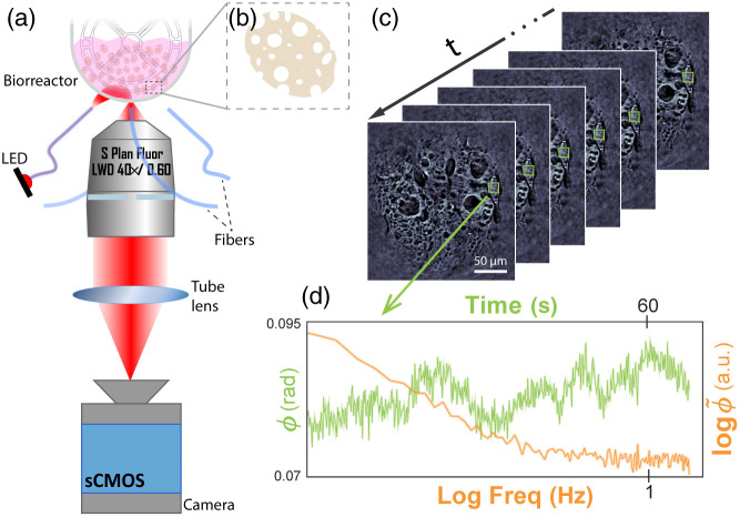
Quantitative oblique back-illumination microscopy (qOBM) is a recently developed label-free imaging technique that enables 3D quantitative phase imaging of thick scattering samples with epi-illumination. Here, we propose dynamic qOBM to achieve functional imaging based on subcellular dynamics, potentially indicative of metabolic activity. We show the potential utility of this novel technique by imaging adherent mesenchymal stromal cells (MSCs) grown in bioreactors, which can help address important unmet needs in cell manufacturing for therapeutics.

AIM

We aim to develop dynamic qOBM and demonstrate its potential for functional imaging based on cellular and subcellular dynamics.

APPROACH

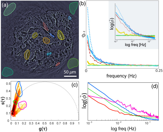
To obtain functional images with dynamic qOBM, a sample is imaged over a period of time and its temporal signals are analyzed. The dynamic signals display an exponential frequency response that can be analyzed with phasor analysis. Functional images of the dynamic signatures are obtained by mapping the frequency dynamic response to phasor space and color-coding clustered signals.

RESULTS

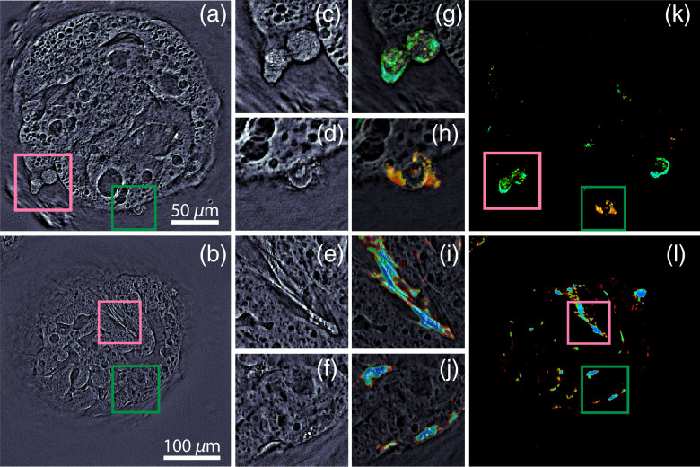
Functional imaging with dynamic qOBM provides unique information related to subcellular activity. The functional qOBM images of MSCs not only improve conspicuity of cells in complex environments (e.g., porous micro-carriers) but also reveal two distinct cell populations with different dynamic behavior.

CONCLUSIONS

In this work we present a label-free, fast, and scalable functional imaging approach to study and intuitively display cellular and subcellular dynamics. We further show the potential utility of this novel technique to help monitor adherent MSCs grown in bioreactors, which can help achieve quality-by-design of cell products, a significant unmet need in the field of cell therapeutics. This approach also has great potential for dynamic studies of other thick samples, such as organoids.

摘要

意义

定量斜向背照明显微镜(qOBM)是一种最近开发的无标记成像技术,可通过 epi 照明对厚散射样品进行 3D 定量相位成像。在这里,我们提出动态 qOBM,以基于亚细胞动力学实现功能成像,这可能表明代谢活性。我们通过对在生物反应器中生长的贴壁间充质基质细胞(MSCs)进行成像来展示这种新技术的潜在应用,这有助于解决细胞治疗中制造方面的一些重要未满足需求。

目的

我们旨在开发动态 qOBM,并证明其基于细胞和亚细胞动力学进行功能成像的潜力。

方法

为了用动态 qOBM 获得功能图像,对样本进行一段时间的成像,并对其时间信号进行分析。动态信号显示出指数频率响应,可以通过相向量分析进行分析。通过将频率动态响应映射到相空间并对聚类信号进行颜色编码,可以获得动态特征的功能图像。

结果

动态 qOBM 的功能成像提供了与亚细胞活性相关的独特信息。MSCs 的功能 qOBM 图像不仅提高了复杂环境(例如多孔微载体)中细胞的对比度,而且还揭示了具有不同动态行为的两种不同细胞群体。

结论

在这项工作中,我们提出了一种无标记、快速且可扩展的功能成像方法,用于研究和直观显示细胞和亚细胞动力学。我们进一步展示了这项新技术的潜在应用,以帮助监测在生物反应器中生长的贴壁 MSCs,这有助于实现细胞产品的质量设计,这是细胞治疗领域的一个重大未满足需求。这种方法对于其他厚样品(如类器官)的动态研究也具有很大的潜力。

https://cdn.ncbi.nlm.nih.gov/pmc/blobs/2614/9243522/1f58cc9ef651/JBO-027-066502-g003.jpg
https://cdn.ncbi.nlm.nih.gov/pmc/blobs/2614/9243522/db555b2a53d2/JBO-027-066502-g001.jpg
https://cdn.ncbi.nlm.nih.gov/pmc/blobs/2614/9243522/4ff10d5cca53/JBO-027-066502-g002.jpg
https://cdn.ncbi.nlm.nih.gov/pmc/blobs/2614/9243522/1f58cc9ef651/JBO-027-066502-g003.jpg
https://cdn.ncbi.nlm.nih.gov/pmc/blobs/2614/9243522/db555b2a53d2/JBO-027-066502-g001.jpg
https://cdn.ncbi.nlm.nih.gov/pmc/blobs/2614/9243522/4ff10d5cca53/JBO-027-066502-g002.jpg
https://cdn.ncbi.nlm.nih.gov/pmc/blobs/2614/9243522/1f58cc9ef651/JBO-027-066502-g003.jpg

相似文献

1
Functional imaging with dynamic quantitative oblique back-illumination microscopy.动态定量反向斜照明显微镜的功能成像。
J Biomed Opt. 2022 Jun;27(6). doi: 10.1117/1.JBO.27.6.066502.
2
Optimization of a flexible fiber-optic probe for epi-mode quantitative phase imaging.优化用于 epi 模式定量相位成像的柔性光纤探头。
Opt Express. 2022 May 23;30(11):17713-17729. doi: 10.1364/OE.454997.
3
Towards in-vivo label-free detection of brain tumor margins with epi-illumination tomographic quantitative phase imaging.用于通过落射照明断层定量相成像进行脑肿瘤边缘的体内无标记检测
Biomed Opt Express. 2021 Feb 25;12(3):1621-1634. doi: 10.1364/BOE.416731. eCollection 2021 Mar 1.
4
GAN-based quantitative oblique back-illumination microscopy enables computationally efficient epi-mode refractive index tomography.基于生成对抗网络的定量斜后照明显微镜技术实现了计算效率高的落射式模式折射率断层扫描。
Biomed Opt Express. 2024 Jul 18;15(8):4764-4774. doi: 10.1364/BOE.528968. eCollection 2024 Aug 1.
5
Quantitative 3D refractive index tomography of opaque samples in epi-mode.落射模式下不透明样品的定量三维折射率断层扫描
Optica. 2021 Jan 20;8(1):6-14. doi: 10.1364/optica.410135.
6
Label-free functional analysis of root-associated microbes with dynamic quantitative oblique back-illumination microscopy.利用动态定量斜向背照显微镜对根相关微生物进行无标记功能分析。
Sci Rep. 2024 Mar 9;14(1):5812. doi: 10.1038/s41598-024-56443-1.
7
Label- and slide-free tissue histology using 3D epi-mode quantitative phase imaging and virtual H&E staining.使用三维落射模式定量相成像和虚拟苏木精-伊红染色的无标记和无切片组织组织学
ArXiv. 2023 Jun 1:arXiv:2306.00548v1.
8
Analysis of structural effects of sickle cell disease on brain vasculature of mice using three-dimensional quantitative phase imaging.利用三维定量相位成像分析镰状细胞病对小鼠脑血管的结构影响。
J Biomed Opt. 2023 Sep;28(9):096501. doi: 10.1117/1.JBO.28.9.096501. Epub 2023 Sep 9.
9
In Vivo Observations of Rapid Scattered Light Changes Associated with Neurophysiological Activity与神经生理活动相关的快速散射光变化的体内观察
10
Noninvasive white blood cell quantification in umbilical cord blood collection bags with quantitative oblique back-illumination microscopy.定量斜向背反射显微镜在脐血采集袋中进行非侵入性白细胞定量。
Transfusion. 2020 Mar;60(3):588-597. doi: 10.1111/trf.15704. Epub 2020 Feb 14.

引用本文的文献

1
Acetic acid enabled nuclear contrast enhancement in epi-mode quantitative phase imaging.乙酸可在落射光模式定量相成像中实现细胞核对比度增强。
J Biomed Opt. 2025 Feb;30(2):026501. doi: 10.1117/1.JBO.30.2.026501. Epub 2025 Feb 4.
2
Non-invasive label-free imaging analysis pipeline for in situ characterization of 3D brain organoids.用于 3D 脑类器官原位特征分析的无创无标记成像分析流程。
Sci Rep. 2024 Sep 27;14(1):22331. doi: 10.1038/s41598-024-72038-2.
3
Non-Invasive Label-free Analysis Pipeline for In Situ Characterization of Differentiation in 3D Brain Organoid Models.

本文引用的文献

1
Three-dimensional dynamics optical coherence tomography for tumor spheroid evaluation.用于肿瘤球体评估的三维动态光学相干断层扫描
Biomed Opt Express. 2021 Oct 12;12(11):6844-6863. doi: 10.1364/BOE.440444. eCollection 2021 Nov 1.
2
Quantitative 3D refractive index tomography of opaque samples in epi-mode.落射模式下不透明样品的定量三维折射率断层扫描
Optica. 2021 Jan 20;8(1):6-14. doi: 10.1364/optica.410135.
3
Clinical and imaging outcomes after intrathecal injection of umbilical cord tissue mesenchymal stem cells in cerebral palsy: a randomized double-blind sham-controlled clinical trial.
用于三维脑类器官模型中分化原位表征的非侵入性无标记分析流程
Res Sq. 2024 Apr 1:rs.3.rs-4049577. doi: 10.21203/rs.3.rs-4049577/v1.
4
Label-free functional analysis of root-associated microbes with dynamic quantitative oblique back-illumination microscopy.利用动态定量斜向背照显微镜对根相关微生物进行无标记功能分析。
Sci Rep. 2024 Mar 9;14(1):5812. doi: 10.1038/s41598-024-56443-1.
5
Label-Free Functional Analysis of Root-Associated Microbes with Dynamic Quantitative Oblique Back-illumination Microscopy.利用动态定量斜后照光显微镜对根际微生物进行无标记功能分析
Res Sq. 2023 Nov 2:rs.3.rs-3517586. doi: 10.21203/rs.3.rs-3517586/v1.
6
Real-time semantic segmentation and anomaly detection of functional images for cell therapy manufacturing.用于细胞治疗生产的功能图像实时语义分割与异常检测
Cytotherapy. 2023 Dec;25(12):1361-1369. doi: 10.1016/j.jcyt.2023.08.011. Epub 2023 Sep 18.
7
Analysis of structural effects of sickle cell disease on brain vasculature of mice using three-dimensional quantitative phase imaging.利用三维定量相位成像分析镰状细胞病对小鼠脑血管的结构影响。
J Biomed Opt. 2023 Sep;28(9):096501. doi: 10.1117/1.JBO.28.9.096501. Epub 2023 Sep 9.
鞘内注射脐带组织间充质干细胞治疗脑瘫的临床和影像学结局:一项随机、双盲、假手术对照临床试验。
Stem Cell Res Ther. 2021 Aug 6;12(1):439. doi: 10.1186/s13287-021-02513-4.
4
Towards in-vivo label-free detection of brain tumor margins with epi-illumination tomographic quantitative phase imaging.用于通过落射照明断层定量相成像进行脑肿瘤边缘的体内无标记检测
Biomed Opt Express. 2021 Feb 25;12(3):1621-1634. doi: 10.1364/BOE.416731. eCollection 2021 Mar 1.
5
Quantitative phase microscopy for non-invasive live cell population monitoring.定量相位显微镜用于非侵入式活细胞群体监测。
Sci Rep. 2021 Feb 24;11(1):4409. doi: 10.1038/s41598-021-83537-x.
6
Dynamic full-field optical coherence tomography: 3D live-imaging of retinal organoids.动态全场光学相干断层扫描:视网膜类器官的三维实时成像
Light Sci Appl. 2020 Aug 17;9:140. doi: 10.1038/s41377-020-00375-8. eCollection 2020.
7
Three dimensional microcarrier system in mesenchymal stem cell culture: a systematic review.间充质干细胞培养中的三维微载体系统:一项系统综述。
Cell Biosci. 2020 Jun 3;10:75. doi: 10.1186/s13578-020-00438-8. eCollection 2020.
8
Fluorescence lifetime imaging microscopy: fundamentals and advances in instrumentation, analysis, and applications.荧光寿命成像显微镜:仪器、分析和应用的基本原理及进展。
J Biomed Opt. 2020 May;25(7):1-43. doi: 10.1117/1.JBO.25.7.071203.
9
Tissue Engineering and Regenerative Medicine: Achievements, Future, and Sustainability in Asia.组织工程与再生医学:亚洲的成就、未来与可持续发展
Front Bioeng Biotechnol. 2020 Mar 24;8:83. doi: 10.3389/fbioe.2020.00083. eCollection 2020.
10
Low-cost fabrication of optical tissue phantoms for use in biomedical imaging.用于生物医学成像的光学组织模型的低成本制造。
Heliyon. 2020 Mar 21;6(3):e03602. doi: 10.1016/j.heliyon.2020.e03602. eCollection 2020 Mar.