Filan Caroline, Charles Seleipiri, Casteleiro Costa Paloma, Niu Weibo, Cheng Brian F, Wen Zhexing, Lu Hang, Robles Francisco E
Georgia Institute of Technology, George W. Woodruff School of Mechanical Engineering, Atlanta, GA, 30318, USA.
Georgia Institute of Technology, Interdisciplinary Program in Bioengineering, Atlanta, GA, 30332, USA.
Res Sq. 2024 Apr 1:rs.3.rs-4049577. doi: 10.21203/rs.3.rs-4049577/v1.
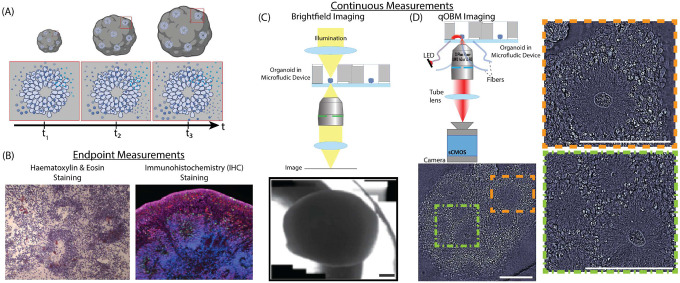
Brain organoids provide a unique opportunity to model organ development in a system similar to human organogenesis . Brain organoids thus hold great promise for drug screening and disease modeling. Conventional approaches to organoid characterization predominantly rely on molecular analysis methods, which are expensive, time-consuming, labor-intensive, and involve the destruction of the valuable 3D architecture of the organoids. This reliance on end-point assays makes it challenging to assess cellular and subcellular events occurring during organoid development in their 3D context. As a result, the long developmental processes are not monitored nor assessed. The ability to perform non-invasive assays is critical for longitudinally assessing features of organoid development during culture. In this paper, we demonstrate a label-free high-content imaging approach for observing changes in organoid morphology and structural changes occurring at the cellular and subcellular level. Enabled by microfluidic-based culture of 3D cell systems and a novel 3D quantitative phase imaging method, we demonstrate the ability to perform non-destructive high-resolution imaging of the organoid. The highlighted results demonstrated in this paper provide a new approach to performing live, non-destructive monitoring of organoid systems during culture.
脑类器官为在类似于人类器官发生的系统中模拟器官发育提供了独特的机会。因此,脑类器官在药物筛选和疾病建模方面具有巨大的潜力。传统的类器官表征方法主要依赖分子分析方法,这些方法昂贵、耗时、劳动强度大,并且会破坏类器官宝贵的三维结构。这种对终点分析的依赖使得在三维环境中评估类器官发育过程中发生的细胞和亚细胞事件具有挑战性。因此,长期的发育过程无法得到监测和评估。进行非侵入性分析的能力对于纵向评估培养过程中类器官发育的特征至关重要。在本文中,我们展示了一种无标记的高内涵成像方法,用于观察类器官形态的变化以及在细胞和亚细胞水平上发生的结构变化。借助基于微流控的三维细胞系统培养和一种新型的三维定量相成像方法,我们展示了对类器官进行无损高分辨率成像的能力。本文突出显示的结果提供了一种在培养过程中对类器官系统进行实时、无损监测的新方法。