• 文献检索
  • 文档翻译
  • 深度研究
  • 学术资讯
  • Suppr Zotero 插件Zotero 插件
  • 邀请有礼
  • 套餐&价格
  • 历史记录
应用&插件
Suppr Zotero 插件Zotero 插件浏览器插件Mac 客户端Windows 客户端微信小程序
定价
高级版会员购买积分包购买API积分包
服务
文献检索文档翻译深度研究API 文档MCP 服务
关于我们
关于 Suppr公司介绍联系我们用户协议隐私条款
关注我们

Suppr 超能文献

核心技术专利:CN118964589B侵权必究
粤ICP备2023148730 号-1Suppr @ 2026

文献检索

告别复杂PubMed语法,用中文像聊天一样搜索,搜遍4000万医学文献。AI智能推荐,让科研检索更轻松。

立即免费搜索

文件翻译

保留排版,准确专业,支持PDF/Word/PPT等文件格式,支持 12+语言互译。

免费翻译文档

深度研究

AI帮你快速写综述,25分钟生成高质量综述,智能提取关键信息,辅助科研写作。

立即免费体验

定量相位显微镜用于非侵入式活细胞群体监测。

Quantitative phase microscopy for non-invasive live cell population monitoring.

机构信息

Phasics, Bâtiment Explorer, Espace Technologique, Route de l'Orme des Merisiers, 91190, St Aubin, France.

Transporter in Imaging and Radiotherapy in Oncology (TIRO), Institut des Sciences et Biotechnologies du Vivant Frédéric Joliot, CEA, School of Medicine, 28 Av de Valombrose, 06107, Nice, France.

出版信息

Sci Rep. 2021 Feb 24;11(1):4409. doi: 10.1038/s41598-021-83537-x.

DOI:10.1038/s41598-021-83537-x
PMID:33627679
原文链接:https://pmc.ncbi.nlm.nih.gov/articles/PMC7904828/
Abstract

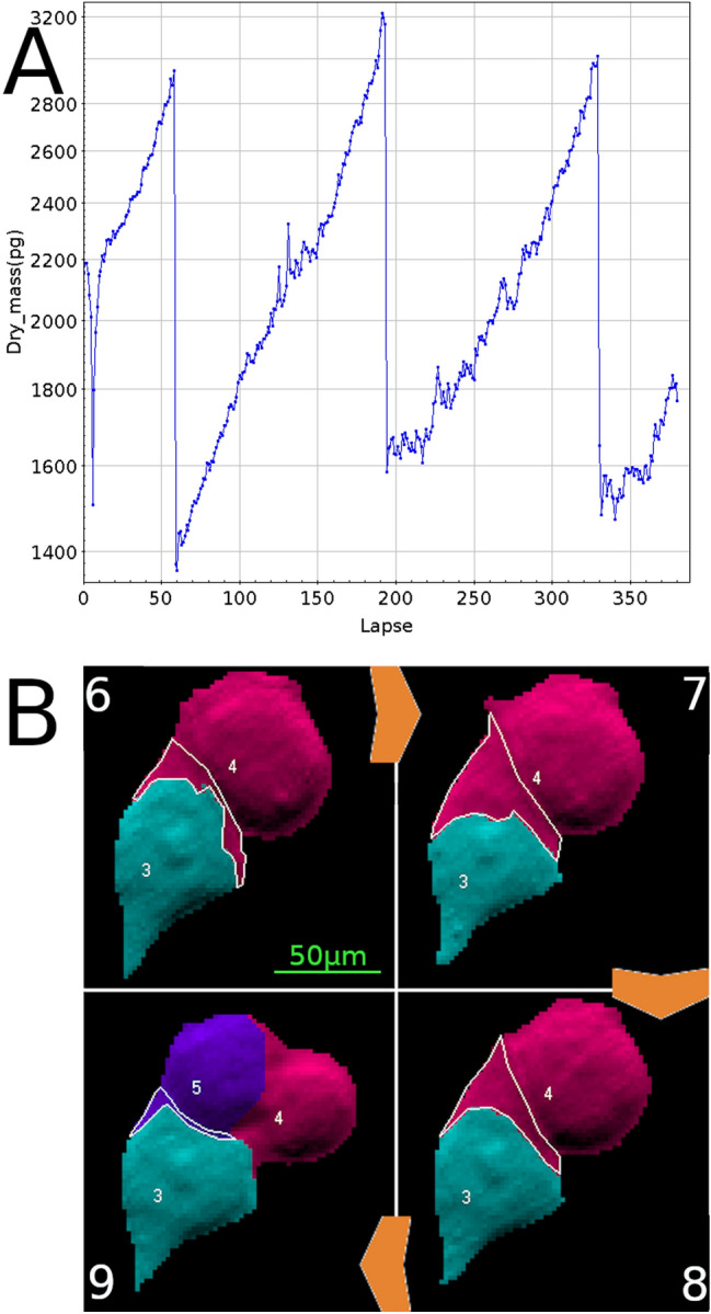
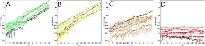
We present here a label-free development based on preexisting Quantitative Phase Imaging (QPI) that allows non-invasive live monitoring of both individual cells and cell populations. Growth, death, effect of toxic compounds are quantified under visible light with a standard inverted microscope. We show that considering the global biomass of a cell population is a more robust and accurate method to assess its growth parameters in comparison to compiling individually segmented cells. This is especially true for confluent conditions. This method expands the use of light microscopy in answering biological questions concerning live cell populations even at high density. In contrast to labeling or lysis of cells this method does not alter the cells and could be useful in high-throughput screening and toxicity studies.

摘要

我们在这里提出了一种基于已有定量相位成像(QPI)的无标记开发,它允许对单个细胞和细胞群体进行非侵入性的实时监测。在可见光下,使用标准倒置显微镜定量测量细胞的生长、死亡和有毒化合物的影响。我们表明,与单独分割细胞相比,考虑细胞群体的整体生物量是评估其生长参数更稳健和准确的方法。对于细胞达到汇合状态时尤其如此。这种方法扩展了使用光学显微镜来回答有关活细胞群体的生物学问题的应用,即使在高细胞密度下也是如此。与细胞标记或裂解不同,该方法不会改变细胞,并且在高通量筛选和毒性研究中可能很有用。

https://cdn.ncbi.nlm.nih.gov/pmc/blobs/3a11/7904828/a8af24bbf648/41598_2021_83537_Fig5_HTML.jpg
https://cdn.ncbi.nlm.nih.gov/pmc/blobs/3a11/7904828/705685d43d23/41598_2021_83537_Fig1_HTML.jpg
https://cdn.ncbi.nlm.nih.gov/pmc/blobs/3a11/7904828/710a1f8fd874/41598_2021_83537_Fig2_HTML.jpg
https://cdn.ncbi.nlm.nih.gov/pmc/blobs/3a11/7904828/86ebb9ddcfc0/41598_2021_83537_Fig3_HTML.jpg
https://cdn.ncbi.nlm.nih.gov/pmc/blobs/3a11/7904828/c37671fe3162/41598_2021_83537_Fig4_HTML.jpg
https://cdn.ncbi.nlm.nih.gov/pmc/blobs/3a11/7904828/a8af24bbf648/41598_2021_83537_Fig5_HTML.jpg

相似文献

1
Quantitative phase microscopy for non-invasive live cell population monitoring.定量相位显微镜用于非侵入式活细胞群体监测。
Sci Rep. 2021 Feb 24;11(1):4409. doi: 10.1038/s41598-021-83537-x.
2
High-throughput, label-free, single-cell, microalgal lipid screening by machine-learning-equipped optofluidic time-stretch quantitative phase microscopy.通过配备机器学习的光流体时间拉伸定量相显微镜进行高通量、无标记、单细胞微藻脂质筛选。
Cytometry A. 2017 May;91(5):494-502. doi: 10.1002/cyto.a.23084. Epub 2017 Apr 11.
3
Multi-ATOM: Ultrahigh-throughput single-cell quantitative phase imaging with subcellular resolution.多原子:具有亚细胞分辨率的超高通量单细胞定量相位成像。
J Biophotonics. 2019 Jul;12(7):e201800479. doi: 10.1002/jbio.201800479. Epub 2019 Apr 1.
4
Quantitative Phase Imaging: Recent Advances and Expanding Potential in Biomedicine.定量相位成像:生物医学中的最新进展和扩展潜力。
ACS Nano. 2022 Aug 23;16(8):11516-11544. doi: 10.1021/acsnano.1c11507. Epub 2022 Aug 2.
5
High-throughput microscopy using live mammalian cells.使用活的哺乳动物细胞的高通量显微镜技术。
Cold Spring Harb Protoc. 2010 Aug 1;2010(7):pdb.top84. doi: 10.1101/pdb.top84.
6
A faster, high resolution, mtPA-GFP-based mitochondrial fusion assay acquiring kinetic data of multiple cells in parallel using confocal microscopy.一种基于mtPA-GFP的更快、高分辨率线粒体融合检测方法,可利用共聚焦显微镜并行获取多个细胞的动力学数据。
J Vis Exp. 2012 Jul 20(65):e3991. doi: 10.3791/3991.
7
Lens-free shadow image based high-throughput continuous cell monitoring technique.无透镜阴影图像的高通量连续细胞监测技术。
Biosens Bioelectron. 2012 Oct-Dec;38(1):126-31. doi: 10.1016/j.bios.2012.05.022. Epub 2012 May 27.
8
A high-throughput three-dimensional cell migration assay for toxicity screening with mobile device-based macroscopic image analysis.高通量三维细胞迁移分析用于基于移动设备的宏观图像分析的毒性筛选。
Sci Rep. 2013 Oct 21;3:3000. doi: 10.1038/srep03000.
9
Advantages of Fresnel biprism-based digital holographic microscopy in quantitative phase imaging.基于菲涅耳双棱镜的数字全息显微镜在定量相位成像中的优势。
J Biomed Opt. 2020 Aug;25(8):1-11. doi: 10.1117/1.JBO.25.8.086501.
10
Dynamic phase differences based on quantitative phase imaging for the objective evaluation of cell behavior.基于定量相成像的动态相位差用于细胞行为的客观评估。
J Biomed Opt. 2015;20(11):111214. doi: 10.1117/1.JBO.20.11.111214.

引用本文的文献

1
Exploring the role of macromolecular crowding and TNFR1 in cell volume control.探讨大分子拥挤和 TNFR1 在细胞体积控制中的作用。
Elife. 2024 Sep 19;13:e92719. doi: 10.7554/eLife.92719.
2
Label-free morpho-molecular phenotyping of living cancer cells by combined Raman spectroscopy and phase tomography.利用拉曼光谱和相衬断层成像技术对活癌细胞进行无标记形态分子表型分析。
Commun Biol. 2024 Jun 29;7(1):785. doi: 10.1038/s42003-024-06496-9.
3
Estimation of the mass density of biological matter from refractive index measurements.从折射率测量估算生物物质的密度。

本文引用的文献

1
Inertial picobalance reveals fast mass fluctuations in mammalian cells.惯性皮重天平揭示了哺乳动物细胞中的快速质量波动。
Nature. 2017 Oct 25;550(7677):500-505. doi: 10.1038/nature24288.
2
Fluorescent proteins such as eGFP lead to catalytic oxidative stress in cells.诸如增强绿色荧光蛋白(eGFP)之类的荧光蛋白会在细胞中引发催化性氧化应激。
Redox Biol. 2017 Aug;12:462-468. doi: 10.1016/j.redox.2017.03.002. Epub 2017 Mar 10.
3
Characterising live cell behaviour: Traditional label-free and quantitative phase imaging approaches.表征活细胞行为:传统的无标记定量相成像方法。
Biophys Rep (N Y). 2024 Jun 12;4(2):100156. doi: 10.1016/j.bpr.2024.100156. Epub 2024 Apr 24.
4
Cell-TypeAnalyzer: A flexible Fiji/ImageJ plugin to classify cells according to user-defined criteria.细胞类型分析器:一个灵活的Fiji/ImageJ插件,可根据用户定义的标准对细胞进行分类。
Biol Imaging. 2022 May 20;2:e5. doi: 10.1017/S2633903X22000058. eCollection 2022.
5
Quantitative phase imaging for characterization of single cell growth dynamics.定量相位成像用于单细胞生长动力学的特征描述。
Lasers Med Sci. 2023 Oct 18;38(1):241. doi: 10.1007/s10103-023-03902-2.
6
Dynamic full-field optical coherence tomography module adapted to commercial microscopes allows longitudinal in vitro cell culture study.经改良适配商用显微镜的动态全场光学相干断层扫描模块可用于体外细胞培养的纵向研究。
Commun Biol. 2023 Sep 28;6(1):992. doi: 10.1038/s42003-023-05378-w.
7
Interface self-referenced dynamic full-field optical coherence tomography.界面自参考动态全场光学相干断层扫描术
Biomed Opt Express. 2023 Jun 21;14(7):3491-3505. doi: 10.1364/BOE.488663. eCollection 2023 Jul 1.
8
Dry mass photometry of single bacteria using quantitative wavefront microscopy.使用定量波前显微镜对单个细菌进行干物质光度测定。
Biophys J. 2023 Aug 8;122(15):3159-3172. doi: 10.1016/j.bpj.2023.06.020. Epub 2023 Jun 30.
9
Low-intensity illumination for lensless digital holographic microscopy with minimized sample interaction.用于无透镜数字全息显微镜的低强度照明,使样品相互作用最小化。
Biomed Opt Express. 2022 Oct 10;13(11):5667-5682. doi: 10.1364/BOE.464367. eCollection 2022 Nov 1.
10
Biomass measurements of single neurites using optical wavefront microscopy.使用光学波前显微镜对单个神经突进行生物量测量。
Biomed Opt Express. 2022 Nov 17;13(12):6550-6560. doi: 10.1364/BOE.471284. eCollection 2022 Dec 1.
Int J Biochem Cell Biol. 2017 Mar;84:89-95. doi: 10.1016/j.biocel.2017.01.004. Epub 2017 Jan 20.
4
Cellular GFP Toxicity and Immunogenicity: Potential Confounders in in Vivo Cell Tracking Experiments.细胞 GFP 毒性和免疫原性:体内细胞示踪实验中的潜在混杂因素。
Stem Cell Rev Rep. 2016 Oct;12(5):553-559. doi: 10.1007/s12015-016-9670-8.
5
Living cell dry mass measurement using quantitative phase imaging with quadriwave lateral shearing interferometry: an accuracy and sensitivity discussion.使用四波横向剪切干涉术的定量相位成像进行活细胞干质量测量:精度与灵敏度探讨
J Biomed Opt. 2015;20(12):126009. doi: 10.1117/1.JBO.20.12.126009.
6
Quantitative retardance imaging of biological samples using quadriwave lateral shearing interferometry.使用四波横向剪切干涉术对生物样品进行定量延迟成像。
Opt Express. 2015 Jun 15;23(12):16383-406. doi: 10.1364/OE.23.016383.
7
Advanced methods of microscope control using μManager software.使用μManager软件的高级显微镜控制方法。
J Biol Methods. 2014;1(2). doi: 10.14440/jbm.2014.36.
8
Live-cell mass profiling: an emerging approach in quantitative biophysics.活细胞质量谱分析:定量生物物理学中的一种新兴方法。
Nat Methods. 2014 Dec;11(12):1221-8. doi: 10.1038/nmeth.3175.
9
Staurosporine analogues from microbial and synthetic sources and their biological activities.微生物和合成来源的星形孢菌素类似物及其生物活性。
Curr Med Chem. 2013;20(31):3872-902. doi: 10.2174/09298673113209990176.
10
Measurement of single cell refractive index, dry mass, volume, and density using a transillumination microscope.使用透照显微镜测量单细胞折射率、干质量、体积和密度。
Phys Rev Lett. 2012 Sep 14;109(11):118105. doi: 10.1103/PhysRevLett.109.118105. Epub 2012 Sep 13.