School of Biological Science and Medical Engineering, Beijing Key Laboratory for Biomaterials and Neural Regeneration, Beijing Advanced Innovation Center for Biomedical Engineering, Beihang University, Beijing, PR China.
Institute of Rehabilitation Engineering, China Rehabilitation Science Institute, Beijing, PR China.
Ann Med. 2022 Dec;54(1):1867-1883. doi: 10.1080/07853890.2022.2089728.
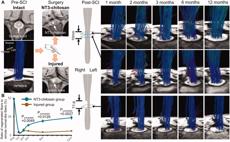
Spinal cord injury (SCI) destroys the sensorimotor pathway and induces brain plasticity. However, the effect of treatment-induced spinal cord tissue regeneration on brain functional reorganization remains unclear. This study was designed to investigate the large-scale functional interactions in the brains of adult female Rhesus monkeys with injured and regenerated thoracic spinal cord.
Resting-state functional magnetic resonance imaging (fMRI) combined with Granger Causality analysis (GCA) and motor behaviour analysis were used to assess the causal interaction between sensorimotor cortices, and calculate the relationship between causal interaction and hindlimb stepping in nine Rhesus monkeys undergoing lesion-induced spontaneous recovery (injured, = 4) and neurotrophin-3/chitosan transplantation-induced regeneration (NT3-chitosan, = 5) after SCI.
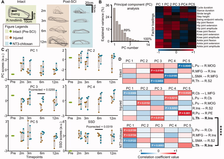
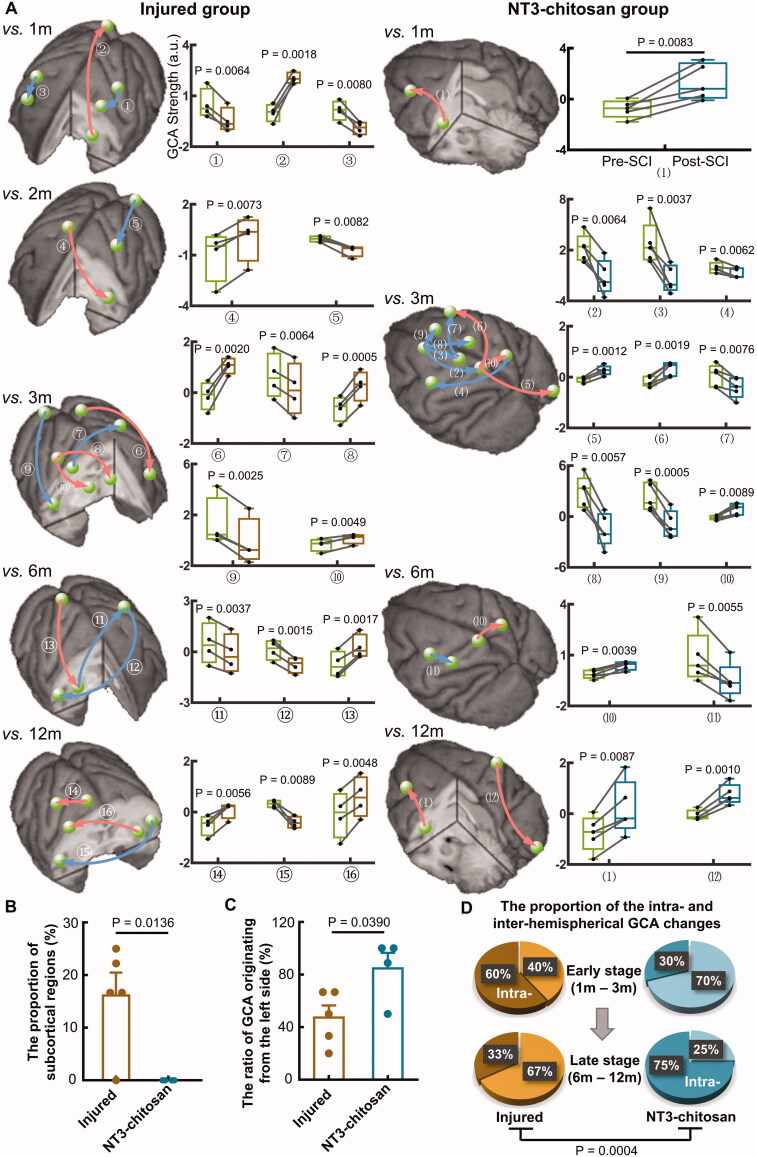
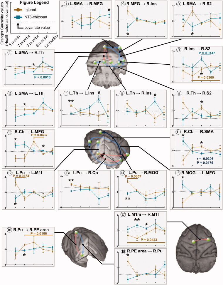
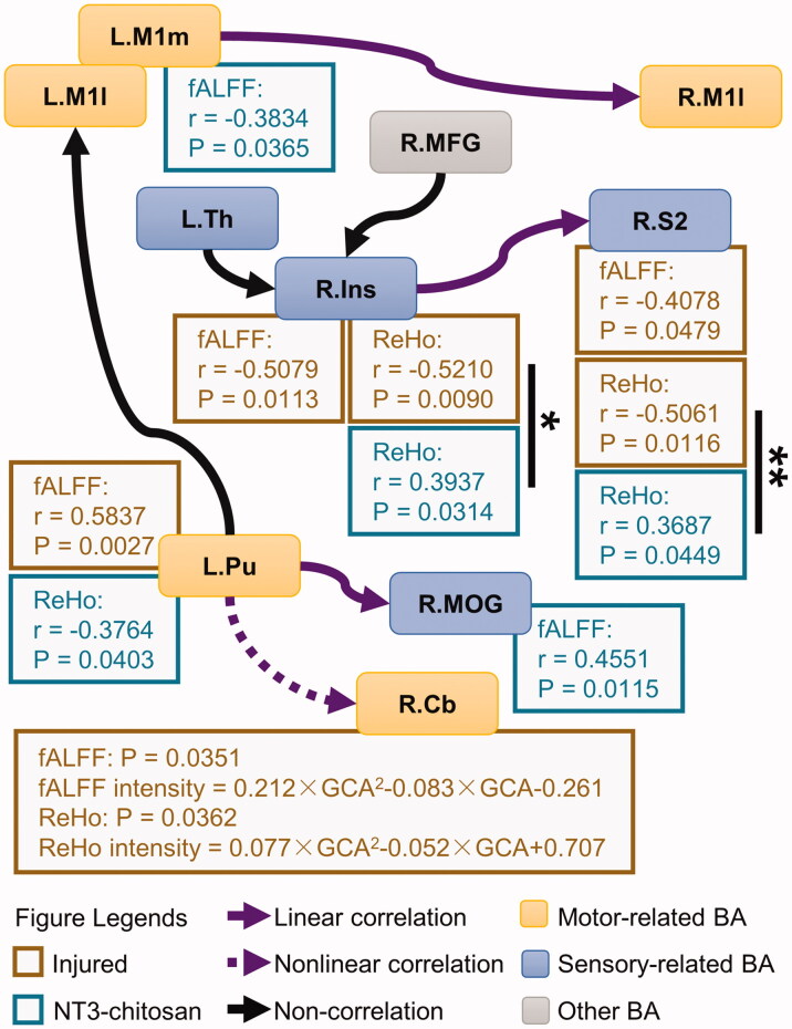
The results showed that the injured and NT3-chitosan-treated animals had distinct spatiotemporal features of brain functional reorganization. The spontaneous recovery followed the model of "early intra-hemispheric reorganization dominant, late inter-hemispheric reorganization dominant", whereas regenerative therapy animals showed the opposite trend. Although the variation degree of information flow intensity was consistent, the tendency and the relationship between local neuronal activity properties and coupling strength were different between the two groups. In addition, the injured and NT3-chitosan-treated animals had similar motor adjustments but various relationship modes between motor performance and information flow intensity.
Our findings show that brain functional reorganization induced by regeneration therapy differed from spontaneous recovery after SCI. The influence of unique changes in brain plasticity on the therapeutic effects of future regeneration therapy strategies should be considered. Key messagesNeural regeneration elicited a unique spatiotemporal mode of brain functional reorganization in the spinal cord injured monkeys, and that regeneration does not simply reverse the process of brain plasticity induced by spinal cord injury (SCI).Independent "properties of local activity - intensity of information flow" relationships between the injured and treated animals indicating that spontaneous recovery and regenerative therapy exerted different effects on the reorganization of the motor network after SCI.A specific information flow from the left thalamus to the right insular can serve as an indicator to reflect a heterogeneous "information flow - motor performance" relationship between injured and treated animals at similar motor adjustments.
脊髓损伤(SCI)破坏了感觉运动通路并诱导了大脑可塑性。然而,治疗诱导的脊髓组织再生对大脑功能重组的影响尚不清楚。本研究旨在探讨损伤和再生胸段脊髓的成年雌性恒河猴大脑中的大规模功能相互作用。
使用静息态功能磁共振成像(fMRI)结合格兰杰因果分析(GCA)和运动行为分析来评估感觉运动皮层之间的因果相互作用,并计算因果相互作用与 9 只恒河猴后肢步进之间的关系在 SCI 后经历损伤诱导的自发恢复(损伤,n=4)和神经营养因子-3/壳聚糖移植诱导的再生(NT3-壳聚糖,n=5)。
结果表明,损伤和 NT3-壳聚糖处理的动物具有明显的大脑功能重组时空特征。自发恢复遵循“早期半球内重组占主导,晚期半球间重组占主导”的模型,而再生治疗动物则呈现相反的趋势。尽管信息流强度的变化程度一致,但两组之间局部神经元活动特性和耦合强度之间的关系和趋势不同。此外,损伤和 NT3-壳聚糖处理的动物具有相似的运动调整,但运动性能和信息流强度之间的关系模式不同。
我们的发现表明,再生治疗诱导的大脑功能重组与 SCI 后的自发恢复不同。应考虑大脑可塑性的独特变化对未来再生治疗策略治疗效果的影响。
神经再生在脊髓损伤的猴子中引起了独特的时空模式的大脑功能重组,并且再生并不能简单地逆转脊髓损伤(SCI)诱导的大脑可塑性过程。
受损和治疗动物之间独立的“局部活动特性-信息流强度”关系表明,自发恢复和再生治疗对 SCI 后运动网络的重组产生了不同的影响。
从左侧丘脑到右侧脑岛的特定信息流可以作为反映受损和治疗动物在相似运动调整下的异质“信息流-运动表现”关系的指标。