Department of Radiology, Shiga University of Medical Science, Seta, Tsukinowa-cho, Otsu, Shiga, 520-2192, Japan.
Department of Radiology, Mayo Clinic, Rochester, MN, USA.
Jpn J Radiol. 2022 Nov;40(11):1105-1120. doi: 10.1007/s11604-022-01305-x. Epub 2022 Jul 9.
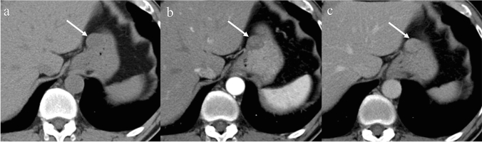
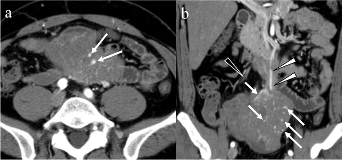
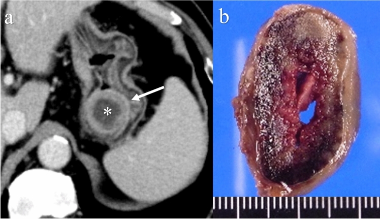
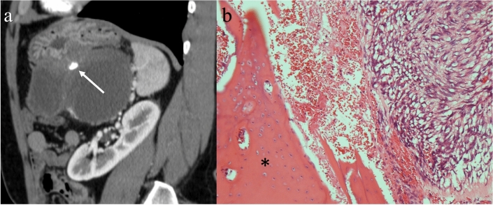
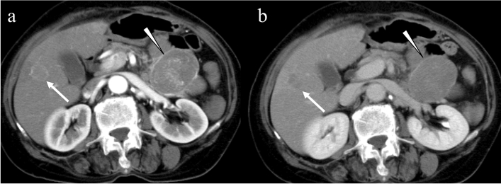
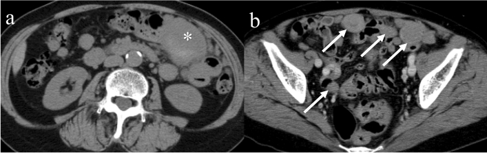
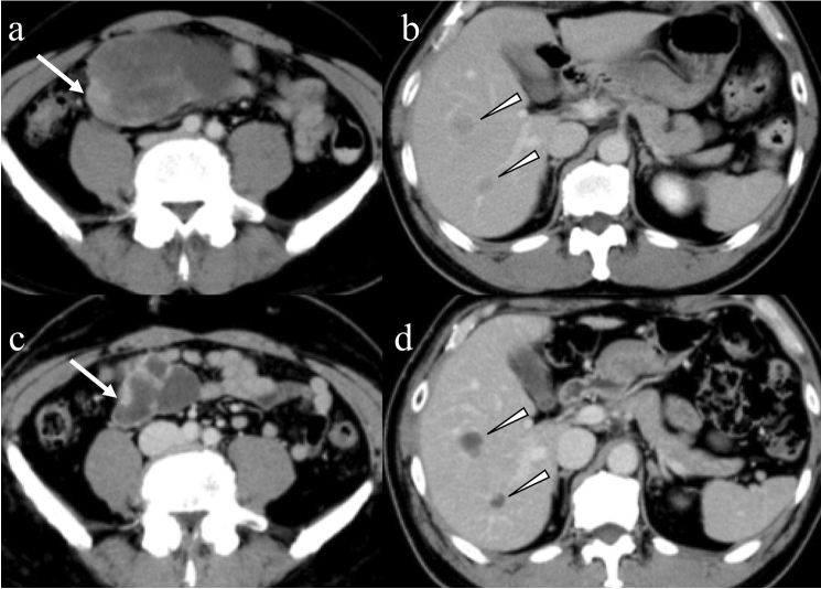
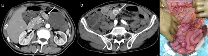
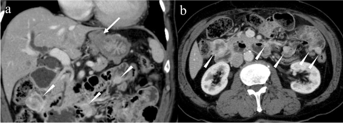
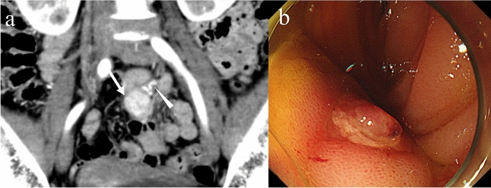
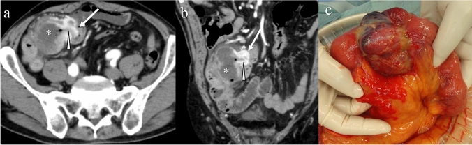
Gastrointestinal stromal tumors (GISTs) originating from the interstitial cells of Cajal in the muscularis propria are the most common mesenchymal tumor of the gastrointestinal tract. Multiple modalities, including computed tomography (CT), magnetic resonance imaging (MRI), fluorodeoxyglucose positron emission tomography, ultrasonography, digital subtraction angiography, and endoscopy, have been performed to evaluate GISTs. CT is most frequently used for diagnosis, staging, surveillance, and response monitoring during molecularly targeted therapy in clinical practice. The diagnosis of GISTs is sometimes challenging because of the diverse imaging findings, such as anatomical location (esophagus, stomach, duodenum, small bowel, colorectum, appendix, and peritoneum), growth pattern, and enhancement pattern as well as the presence of necrosis, calcification, ulceration, early venous return, and metastasis. Imaging findings of GISTs treated with antineoplastic agents are quite different from those of other neoplasms (e.g. adenocarcinomas) because only subtle changes in size are seen even in responsive lesions. Furthermore, the recurrence pattern of GISTs is different from that of other neoplasms. This review discusses the advantages and disadvantages of each imaging modality, describes imaging findings obtained before and after treatment, presents a few cases of complicated GISTs, and discusses recent investigations performed using CT and MRI to predict histological risk grade, gene mutations, and patient outcomes.
胃肠道间质瘤(GISTs)起源于固有肌层的 Cajal 间质细胞,是胃肠道最常见的间叶性肿瘤。多种影像学模态,包括计算机断层扫描(CT)、磁共振成像(MRI)、氟脱氧葡萄糖正电子发射断层扫描、超声、数字减影血管造影和内镜,已被用于评估 GISTs。在临床实践中,CT 最常用于诊断、分期、监测和分子靶向治疗期间的反应监测。由于 GISTs 的影像学表现多样,例如解剖位置(食管、胃、十二指肠、小肠、结直肠、阑尾和腹膜)、生长模式和增强模式以及坏死、钙化、溃疡、早期静脉回流和转移的存在,其诊断有时具有挑战性。接受抗肿瘤药物治疗的 GISTs 的影像学表现与其他肿瘤(例如腺癌)明显不同,因为即使是在有反应的病变中,也只能看到大小的细微变化。此外,GISTs 的复发模式与其他肿瘤不同。本文讨论了每种影像学模态的优缺点,描述了治疗前后的影像学表现,展示了一些复杂 GIST 的病例,并讨论了使用 CT 和 MRI 进行的预测组织学风险分级、基因突变和患者预后的最新研究。