Department of Radiology, Peking University First Hospital, Peking University, Beijing, China.
Department of General Surgery, Peking University First Hospital, Peking University, Beijing, China.
Korean J Radiol. 2021 Mar;22(3):344-353. doi: 10.3348/kjr.2019.0851. Epub 2020 Oct 21.
The mitotic count of gastrointestinal stromal tumors (GIST) is closely associated with the risk of planting and metastasis. The purpose of this study was to develop a predictive model for the mitotic index of local primary GIST, based on deep learning algorithm.
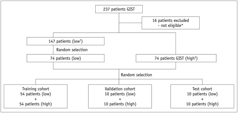
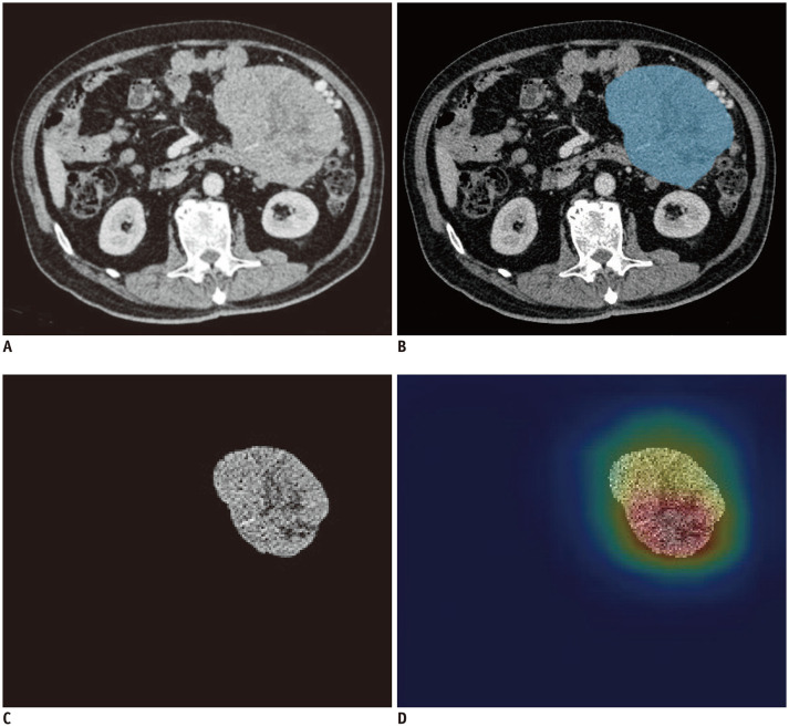
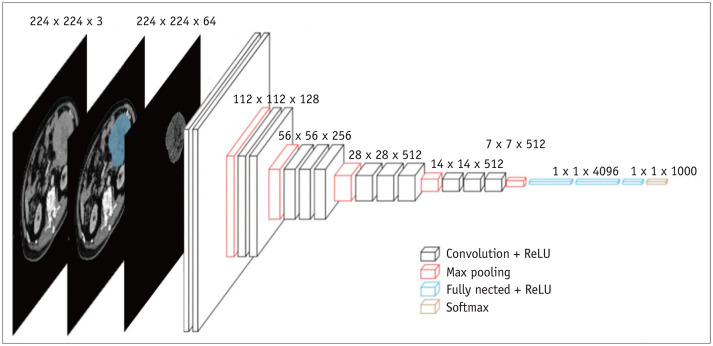
Abdominal contrast-enhanced CT images of 148 pathologically confirmed GIST cases were retrospectively collected for the development of a deep learning classification algorithm. The areas of GIST masses on the CT images were retrospectively labelled by an experienced radiologist. The postoperative pathological mitotic count was considered as the gold standard (high mitotic count, > 5/50 high-power fields [HPFs]; low mitotic count, ≤ 5/50 HPFs). A binary classification model was trained on the basis of the VGG16 convolutional neural network, using the CT images with the training set (n = 108), validation set (n = 20), and the test set (n = 20). The sensitivity, specificity, positive predictive value (PPV), and negative predictive value (NPV) were calculated at both, the image level and the patient level. The receiver operating characteristic curves were generated on the basis of the model prediction results and the area under curves (AUCs) were calculated. The risk categories of the tumors were predicted according to the Armed Forces Institute of Pathology criteria.
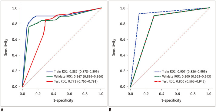
At the image level, the classification prediction results of the mitotic counts in the test cohort were as follows: sensitivity 85.7% (95% confidence interval [CI]: 0.834-0.877), specificity 67.5% (95% CI: 0.636-0.712), PPV 82.1% (95% CI: 0.797-0.843), NPV 73.0% (95% CI: 0.691-0.766), and AUC 0.771 (95% CI: 0.750-0.791). At the patient level, the classification prediction results in the test cohort were as follows: sensitivity 90.0% (95% CI: 0.541-0.995), specificity 70.0% (95% CI: 0.354-0.919), PPV 75.0% (95% CI: 0.428-0.933), NPV 87.5% (95% CI: 0.467-0.993), and AUC 0.800 (95% CI: 0.563-0.943).
We developed and preliminarily verified the GIST mitotic count binary prediction model, based on the VGG convolutional neural network. The model displayed a good predictive performance.
胃肠道间质瘤(GIST)的有丝分裂计数与种植和转移的风险密切相关。本研究旨在基于深度学习算法,为局部原发性 GIST 的有丝分裂指数建立预测模型。
回顾性收集了 148 例经病理证实的 GIST 病例的腹部增强 CT 图像,用于开发深度学习分类算法。由一名有经验的放射科医生对 CT 图像上的 GIST 肿块区域进行回顾性标记。术后病理有丝分裂计数被视为金标准(高有丝分裂计数,> 5/50 高倍视野[HPF];低有丝分裂计数,≤ 5/50 HPF)。基于 VGG16 卷积神经网络,使用训练集(n = 108)、验证集(n = 20)和测试集(n = 20)对 CT 图像进行了二进制分类模型训练。计算了图像和患者水平的灵敏度、特异性、阳性预测值(PPV)和阴性预测值(NPV)。根据模型预测结果生成受试者工作特征曲线,并计算曲线下面积(AUC)。根据武装部队病理研究所的标准预测肿瘤的危险类别。
在图像水平上,测试队列中核分裂计数的分类预测结果如下:灵敏度 85.7%(95%置信区间[CI]:0.834-0.877),特异性 67.5%(95%CI:0.636-0.712),PPV 82.1%(95%CI:0.797-0.843),NPV 73.0%(95%CI:0.691-0.766),AUC 0.771(95%CI:0.750-0.791)。在患者水平上,测试队列中的分类预测结果如下:灵敏度 90.0%(95%CI:0.541-0.995),特异性 70.0%(95%CI:0.354-0.919),PPV 75.0%(95%CI:0.428-0.933),NPV 87.5%(95%CI:0.467-0.993),AUC 0.800(95%CI:0.563-0.943)。
我们基于 VGG 卷积神经网络开发并初步验证了 GIST 有丝分裂计数的二元预测模型。该模型表现出良好的预测性能。