Angom Ramcharan Singh, Kulkarni Tanmay, Wang Enfeng, Kumar Dutta Shamit, Bhattacharya Santanu, Das Pritam, Mukhopadhyay Debabrata
Department of Biochemistry and Molecular Biology, Jacksonville, FL, United States.
Department of Physiology and Biomedical Engineering, Mayo Clinic College of Medicine and Science, Jacksonville, FL, United States.
Front Cell Dev Biol. 2022 Jul 1;10:903047. doi: 10.3389/fcell.2022.903047. eCollection 2022.
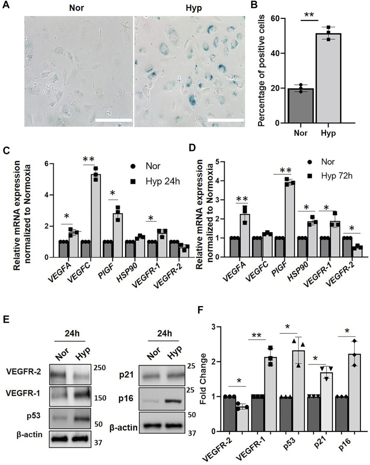
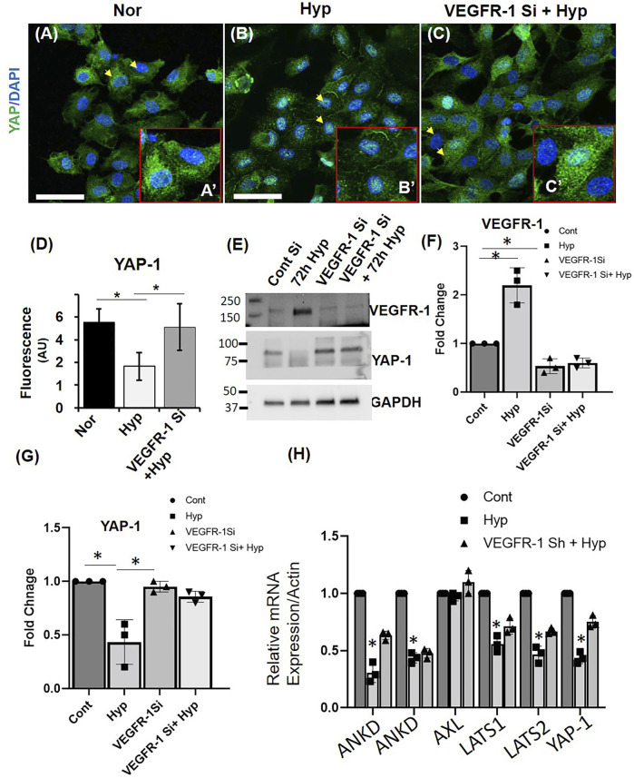
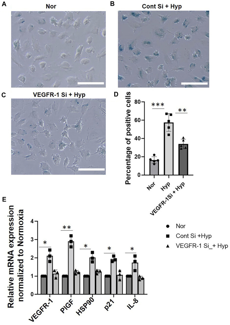
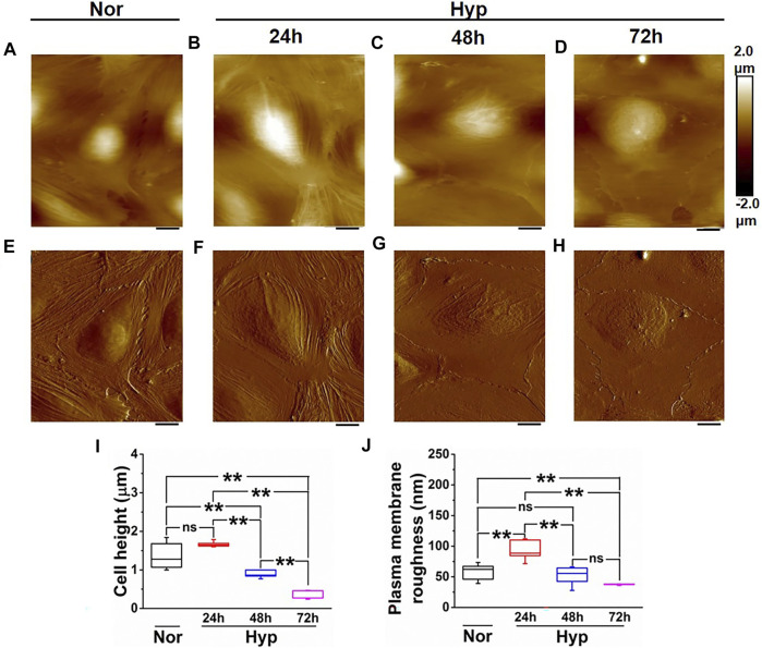
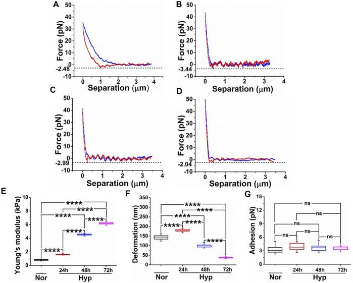
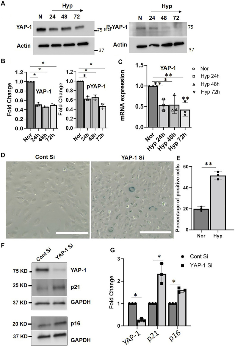
Hypoxia-induced endothelial cell (EC) dysfunction has been implicated as potential initiators of different pathogenesis, including Alzheimer's disease and vascular dementia. However, in-depth structural, mechanical, and molecular mechanisms leading to EC dysfunction and pathology need to be revealed. Here, we show that ECs exposed to hypoxic conditions readily enter a senescence phenotype. As expected, hypoxia upregulated the expression of vascular endothelial growth factor (VEGFs) and its receptors (VEGFRs) in the ECs. Interestingly, Knockdown of VEGFR-1 expression prior to hypoxia exposure prevented EC senescence, suggesting an important role of VEGFR-1 expression in the induction of EC senescence. Using atomic force microscopy, we showed that senescent ECs had a flattened cell morphology, decreased membrane ruffling, and increased membrane stiffness, demonstrating unique morphological and nanomechanical signatures. Furthermore, we show that hypoxia inhibited the Hippo pathway Yes-associated protein (YAP-1) expression and knockdown of YAP-1 induced senescence in the ECs, supporting a key role of YAP-1 expression in the induction of EC senescence. And importantly, VEGFR-1 Knockdown in the ECs modulated YAP-1 expression, suggesting a novel VEGFR-1-YAP-1 axis in the induction of hypoxia-mediated EC senescence. In conclusion, VEGFR-1 is overexpressed in ECs undergoing hypoxia-mediated senescence, and the knockdown of VEGFR-1 restores cellular structural and nanomechanical integrity by recovering YAP-1 expression.
缺氧诱导的内皮细胞(EC)功能障碍被认为是包括阿尔茨海默病和血管性痴呆在内的不同发病机制的潜在启动因素。然而,导致EC功能障碍和病理的深入结构、机械和分子机制仍有待揭示。在此,我们表明暴露于缺氧条件下的EC很容易进入衰老表型。正如预期的那样,缺氧上调了EC中血管内皮生长因子(VEGFs)及其受体(VEGFRs)的表达。有趣的是,在缺氧暴露前敲低VEGFR-1的表达可防止EC衰老,这表明VEGFR-1表达在诱导EC衰老中起重要作用。使用原子力显微镜,我们发现衰老的EC具有扁平的细胞形态、减少的膜褶皱和增加的膜硬度,表现出独特的形态和纳米力学特征。此外,我们表明缺氧抑制了Hippo通路Yes相关蛋白(YAP-1)的表达,敲低YAP-1可诱导EC衰老,这支持了YAP-1表达在诱导EC衰老中的关键作用。重要的是,在EC中敲低VEGFR-1可调节YAP-1的表达,这表明在缺氧介导的EC衰老诱导中存在一种新的VEGFR-1-YAP-1轴。总之,VEGFR-1在经历缺氧介导衰老的EC中过表达,敲低VEGFR-1可通过恢复YAP-1表达来恢复细胞结构和纳米力学完整性。