Samson Genelle D, Lahti Adrienne C, Kraguljac Nina V
Department of Psychology, University of Alabama at Birmingham, Birmingham, AL, USA.
Department of Psychiatry and Behavioral Neurobiology, University of Alabama at Birmingham, Birmingham, AL, USA.
Schizophrenia (Heidelb). 2022 Apr 26;8(1):42. doi: 10.1038/s41537-022-00245-9.
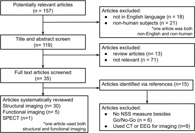
Neurological soft signs (NSS) are common in patients with schizophrenia. However, the neural substrates of NSS remain poorly understood. Using legacy PubMed, we performed a systematic review and included studies that assessed NSS and obtained neuroimaging data in patients with a schizophrenia spectrum disorder published up to June 2020. We systematically reviewed 35 relevant articles. Studies consistently implicate the basal ganglia and cerebellum as structural substrates of NSS and suggest that somatomotor and somatosensory regions as well as areas involved in visual processing and spatial orientation may underlie NSS in psychosis spectrum disorders. Additionally, dysfunction of frontoparietal and cerebellar networks has been implicated in the pathophysiology of NSS. The current literature outlines several structural and functional brain signatures that are relevant for NSS in schizophrenia spectrum disorder. The majority of studies assessed gray matter structure, but only a few studies leveraged other imaging methods such as diffusion weighted imaging, or molecular imaging. Due to this, it remains unclear if white matter integrity deficits or neurometabolic alterations contribute to NSS in the illness. While a substantial portion of the literature has been conducted in patients in the early illness stages, mitigating confounds of illness chronicity, few studies have been conducted in antipsychotic medication-naïve patients, which is a clear limitation. Furthermore, only little is known about the temporal evolution of NSS and associated brain signatures. Future studies addressing these pivotal gaps in our mechanistic understanding of NSS will be important.
神经软体征(NSS)在精神分裂症患者中很常见。然而,NSS的神经基质仍知之甚少。我们使用传统的PubMed进行了一项系统综述,纳入了截至2020年6月发表的评估NSS并获取精神分裂症谱系障碍患者神经影像学数据的研究。我们系统地回顾了35篇相关文章。研究一致表明基底神经节和小脑是NSS的结构基质,并表明躯体运动和躯体感觉区域以及参与视觉处理和空间定向的区域可能是精神病谱系障碍中NSS的基础。此外,额顶叶和小脑网络功能障碍与NSS的病理生理学有关。当前文献概述了几种与精神分裂症谱系障碍中NSS相关的脑结构和功能特征。大多数研究评估了灰质结构,但只有少数研究利用了其他成像方法,如扩散加权成像或分子成像。因此,尚不清楚白质完整性缺陷或神经代谢改变是否导致该疾病中的NSS。虽然大部分文献是在疾病早期阶段的患者中进行的,以减轻疾病慢性化的混杂因素,但很少有研究是在未使用抗精神病药物的患者中进行的,这是一个明显的局限性。此外,关于NSS及其相关脑特征的时间演变知之甚少。未来针对我们对NSS机制理解中的这些关键差距进行的研究将很重要。