• 文献检索
  • 文档翻译
  • 深度研究
  • 学术资讯
  • Suppr Zotero 插件Zotero 插件
  • 邀请有礼
  • 套餐&价格
  • 历史记录
应用&插件
Suppr Zotero 插件Zotero 插件浏览器插件Mac 客户端Windows 客户端微信小程序
定价
高级版会员购买积分包购买API积分包
服务
文献检索文档翻译深度研究API 文档MCP 服务
关于我们
关于 Suppr公司介绍联系我们用户协议隐私条款
关注我们

Suppr 超能文献

核心技术专利:CN118964589B侵权必究
粤ICP备2023148730 号-1Suppr @ 2026

文献检索

告别复杂PubMed语法,用中文像聊天一样搜索,搜遍4000万医学文献。AI智能推荐,让科研检索更轻松。

立即免费搜索

文件翻译

保留排版,准确专业,支持PDF/Word/PPT等文件格式,支持 12+语言互译。

免费翻译文档

深度研究

AI帮你快速写综述,25分钟生成高质量综述,智能提取关键信息,辅助科研写作。

立即免费体验

基于多重分形方法的乳腺 ROI 内微钙化检测分类。

Classification of Mammographic ROI for Microcalcification Detection Using Multifractal Approach.

机构信息

Biomedical Engineering Laboratory, Faculty of Technology, Abou Bekr Belkaid University, 13000, Tlemcen, Algeria.

出版信息

J Digit Imaging. 2022 Dec;35(6):1544-1559. doi: 10.1007/s10278-022-00677-w. Epub 2022 Jul 19.

DOI:10.1007/s10278-022-00677-w
PMID:35854037
原文链接:https://pmc.ncbi.nlm.nih.gov/articles/PMC9712886/
Abstract

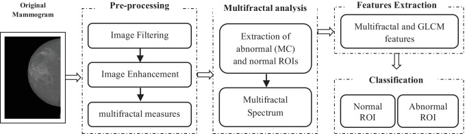
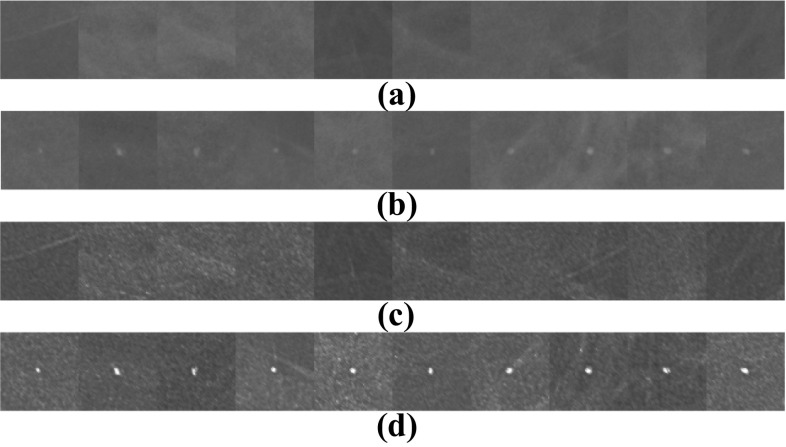
Microcalcifications (MCs) are the main signs of precancerous cells. The development of aided-system for their detection has become a challenge for researchers in this field. In this paper, we propose a system for MCs detection based on the multifractal approach that classifies mammographic ROIs into normal (healthy) or abnormal ROIs containing MCs. The proposed method is divided into four main steps: a mammogram pre-processing step based on breast selection, breast density reduction using haze removal algorithm and contrast enhancement using multifractal measures. The second step consists of extracting the normal and abnormal ROIs and calculating the multifractal spectrum of each ROI. The next step represents the extraction of the multifractal features from the multifractal spectrum and the GLCM characteristics of each ROI. The last step is the classification of ROIs where three classifiers are tested (KNN, DT, and SVM). The system is evaluated on images from the INbreast database (308 images) with a total of 2688 extracted ROIs (1344 normal, 1344 with MC) from different BI-RADS classes. In this study, the SVM classifier gave the best classification results with a sensitivity, specificity, and precision of 98.66%, 97.77%, and 98.20% respectively. These results are very satisfactory and remarkable compared to the literature.

摘要

微钙化(MCs)是癌前细胞的主要特征。开发辅助系统来检测它们已成为该领域研究人员的一项挑战。在本文中,我们提出了一种基于多重分形方法的 MCs 检测系统,该系统将乳腺 ROI 分为正常(健康)或包含 MCs 的异常 ROI。所提出的方法分为四个主要步骤:基于乳腺选择的乳腺预处理步骤、使用去雾算法降低乳腺密度和使用多重分形度量进行对比度增强。第二步包括提取正常和异常 ROI,并计算每个 ROI 的多重分形谱。第三步表示从多重分形谱和每个 ROI 的 GLCM 特征中提取多重分形特征。最后一步是对 ROI 进行分类,测试了三种分类器(KNN、DT 和 SVM)。该系统在 INbreast 数据库(308 张图像)上进行了评估,共从不同 BI-RADS 类别中提取了 2688 个 ROI(1344 个正常,1344 个有 MC)。在这项研究中,SVM 分类器的分类效果最好,其灵敏度、特异性和精度分别为 98.66%、97.77%和 98.20%。与文献相比,这些结果非常令人满意和显著。

https://cdn.ncbi.nlm.nih.gov/pmc/blobs/2f92/9712886/d8538ce1dc0b/10278_2022_677_Fig10_HTML.jpg
https://cdn.ncbi.nlm.nih.gov/pmc/blobs/2f92/9712886/30168ab74285/10278_2022_677_Fig1_HTML.jpg
https://cdn.ncbi.nlm.nih.gov/pmc/blobs/2f92/9712886/1e20b61f09d3/10278_2022_677_Fig2_HTML.jpg
https://cdn.ncbi.nlm.nih.gov/pmc/blobs/2f92/9712886/43c8b67ed9fe/10278_2022_677_Fig3_HTML.jpg
https://cdn.ncbi.nlm.nih.gov/pmc/blobs/2f92/9712886/487eb0918fb2/10278_2022_677_Fig4_HTML.jpg
https://cdn.ncbi.nlm.nih.gov/pmc/blobs/2f92/9712886/19749f0ecdcc/10278_2022_677_Fig5_HTML.jpg
https://cdn.ncbi.nlm.nih.gov/pmc/blobs/2f92/9712886/b34e9b426d3c/10278_2022_677_Fig6_HTML.jpg
https://cdn.ncbi.nlm.nih.gov/pmc/blobs/2f92/9712886/50b43724bbfa/10278_2022_677_Fig7_HTML.jpg
https://cdn.ncbi.nlm.nih.gov/pmc/blobs/2f92/9712886/7f13e47943fd/10278_2022_677_Fig8_HTML.jpg
https://cdn.ncbi.nlm.nih.gov/pmc/blobs/2f92/9712886/069a6891a4c9/10278_2022_677_Fig9_HTML.jpg
https://cdn.ncbi.nlm.nih.gov/pmc/blobs/2f92/9712886/d8538ce1dc0b/10278_2022_677_Fig10_HTML.jpg

相似文献

1
Classification of Mammographic ROI for Microcalcification Detection Using Multifractal Approach.基于多重分形方法的乳腺 ROI 内微钙化检测分类。
J Digit Imaging. 2022 Dec;35(6):1544-1559. doi: 10.1007/s10278-022-00677-w. Epub 2022 Jul 19.
2
Breast microcalcifications detection based on fusing features with DTCWT.基于 DTCWT 融合特征的乳腺微钙化检测。
J Xray Sci Technol. 2020;28(2):197-218. doi: 10.3233/XST-190583.
3
False Positive Reduction by an Annular Model as a Set of Few Features for Microcalcification Detection to Assist Early Diagnosis of Breast Cancer.环状模型减少微钙化检测中的假阳性作为少量特征集,辅助乳腺癌早期诊断。
J Med Syst. 2018 Jun 18;42(8):134. doi: 10.1007/s10916-018-0989-3.
4
A novel machine learning approach on texture analysis for automatic breast microcalcification diagnosis classification of mammogram images.一种基于纹理分析的新型机器学习方法,用于自动乳腺微钙化诊断分类的乳腺 X 线图像。
J Cancer Res Clin Oncol. 2023 Aug;149(9):6151-6170. doi: 10.1007/s00432-023-04571-y. Epub 2023 Jan 21.
5
Fissures segmentation using surface features: content-based retrieval for mammographic mass using ensemble classifier.利用表面特征进行裂隙分割:基于内容的乳腺肿块检索使用集成分类器。
Acad Radiol. 2011 Dec;18(12):1475-84. doi: 10.1016/j.acra.2011.08.012.
6
Evaluating geodesic active contours in microcalcifications segmentation on mammograms.评估乳腺钼靶片中微钙化分割的测地主动轮廓。
Comput Methods Programs Biomed. 2015 Dec;122(3):304-15. doi: 10.1016/j.cmpb.2015.08.016. Epub 2015 Aug 29.
7
Automatic detection of clustered microcalcifications in digital mammograms: Study on applying adaboost with SVM-based component classifiers.数字乳腺X线摄影中簇状微钙化的自动检测:基于支持向量机的组件分类器应用Adaboost算法的研究
Annu Int Conf IEEE Eng Med Biol Soc. 2008;2008:4789-92. doi: 10.1109/IEMBS.2008.4650284.
8
SVM based system for classification of microcalcifications in digital mammograms.基于支持向量机的数字乳腺X线摄影中微钙化分类系统。
Conf Proc IEEE Eng Med Biol Soc. 2006;2006:4747-50. doi: 10.1109/IEMBS.2006.259320.
9
Pattern Recognition and Size Prediction of Microcalcification Based on Physical Characteristics by Using Digital Mammogram Images.基于数字乳腺图像物理特征的微钙化模式识别与大小预测。
J Digit Imaging. 2018 Dec;31(6):912-922. doi: 10.1007/s10278-018-0075-x.
10
Quantitative comparison of clustered microcalcifications in for-presentation and for-processing mammograms in full-field digital mammography.全数字化乳腺摄影中用于诊断和存档的钼靶片中小钙化簇的定量比较。
Med Phys. 2017 Jul;44(7):3726-3738. doi: 10.1002/mp.12316. Epub 2017 Jun 9.

本文引用的文献

1
A machine learning approach on multiscale texture analysis for breast microcalcification diagnosis.基于多尺度纹理分析的机器学习方法在乳腺微钙化诊断中的应用。
BMC Bioinformatics. 2020 Mar 11;21(Suppl 2):91. doi: 10.1186/s12859-020-3358-4.
2
Classification of Mammogram Images Using Multiscale all Convolutional Neural Network (MA-CNN).使用多尺度全卷积神经网络(MA-CNN)对乳腺 X 光图像进行分类。
J Med Syst. 2019 Dec 14;44(1):30. doi: 10.1007/s10916-019-1494-z.
3
A Hybridized ELM for Automatic Micro Calcification Detection in Mammogram Images Based on Multi-Scale Features.
基于多尺度特征的乳腺钼钯图像中自动微钙化检测的混合 ELM
J Med Syst. 2019 May 15;43(7):183. doi: 10.1007/s10916-019-1316-3.
4
Pattern Recognition and Size Prediction of Microcalcification Based on Physical Characteristics by Using Digital Mammogram Images.基于数字乳腺图像物理特征的微钙化模式识别与大小预测。
J Digit Imaging. 2018 Dec;31(6):912-922. doi: 10.1007/s10278-018-0075-x.
5
Classification of micro-calcification in mammograms using scalable linear Fisher discriminant analysis.使用可扩展线性 Fisher 判别分析对乳腺 X 线片中的微钙化进行分类。
Med Biol Eng Comput. 2018 Aug;56(8):1475-1485. doi: 10.1007/s11517-017-1774-z. Epub 2018 Jan 25.
6
Applications of Support Vector Machine (SVM) Learning in Cancer Genomics.支持向量机(SVM)学习在癌症基因组学中的应用。
Cancer Genomics Proteomics. 2018 Jan-Feb;15(1):41-51. doi: 10.21873/cgp.20063.
7
Multiscale multifractal detrended-fluctuation analysis of two-dimensional surfaces.二维表面的多尺度多重分形去趋势波动分析。
Phys Rev E. 2016 Apr;93:042213. doi: 10.1103/PhysRevE.93.042213. Epub 2016 Apr 21.
8
INbreast: toward a full-field digital mammographic database.INbreast:迈向全视野数字化乳腺 X 光摄影数据库。
Acad Radiol. 2012 Feb;19(2):236-48. doi: 10.1016/j.acra.2011.09.014. Epub 2011 Nov 10.
9
Fractal and multifractal analysis: a review.分形与多重分形分析:综述
Med Image Anal. 2009 Aug;13(4):634-49. doi: 10.1016/j.media.2009.05.003. Epub 2009 May 27.
10
Direct determination of the f( alpha ) singularity spectrum.f(α)奇异性谱的直接测定。
Phys Rev Lett. 1989 Mar 20;62(12):1327-1330. doi: 10.1103/PhysRevLett.62.1327.