• 文献检索
  • 文档翻译
  • 深度研究
  • 学术资讯
  • Suppr Zotero 插件Zotero 插件
  • 邀请有礼
  • 套餐&价格
  • 历史记录
应用&插件
Suppr Zotero 插件Zotero 插件浏览器插件Mac 客户端Windows 客户端微信小程序
定价
高级版会员购买积分包购买API积分包
服务
文献检索文档翻译深度研究API 文档MCP 服务
关于我们
关于 Suppr公司介绍联系我们用户协议隐私条款
关注我们

Suppr 超能文献

核心技术专利:CN118964589B侵权必究
粤ICP备2023148730 号-1Suppr @ 2026

文献检索

告别复杂PubMed语法,用中文像聊天一样搜索,搜遍4000万医学文献。AI智能推荐,让科研检索更轻松。

立即免费搜索

文件翻译

保留排版,准确专业,支持PDF/Word/PPT等文件格式,支持 12+语言互译。

免费翻译文档

深度研究

AI帮你快速写综述,25分钟生成高质量综述,智能提取关键信息,辅助科研写作。

立即免费体验

基于多尺度纹理分析的机器学习方法在乳腺微钙化诊断中的应用。

A machine learning approach on multiscale texture analysis for breast microcalcification diagnosis.

机构信息

I.R.C.C.S. Istituto Tumori "Giovanni Paolo II", viale O. Flacco 65, Bari, Italy.

Dip. Interateneo di Fisica "M. Merlin", Università degli Studi di Bari "A. Moro", via G. Amendola 173, Bari, Italy.

出版信息

BMC Bioinformatics. 2020 Mar 11;21(Suppl 2):91. doi: 10.1186/s12859-020-3358-4.

DOI:10.1186/s12859-020-3358-4
PMID:32164532
原文链接:https://pmc.ncbi.nlm.nih.gov/articles/PMC7069158/
Abstract

BACKGROUND

Screening programs use mammography as primary diagnostic tool for detecting breast cancer at an early stage. The diagnosis of some lesions, such as microcalcifications, is still difficult today for radiologists. In this paper, we proposed an automatic binary model for discriminating tissue in digital mammograms, as support tool for the radiologists. In particular, we compared the contribution of different methods on the feature selection process in terms of the learning performances and selected features.

RESULTS

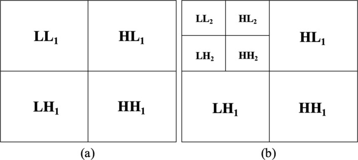
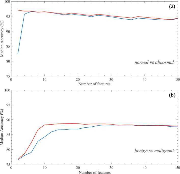
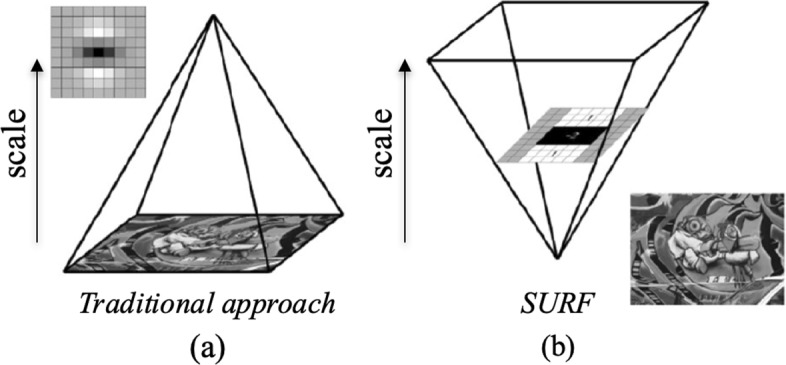
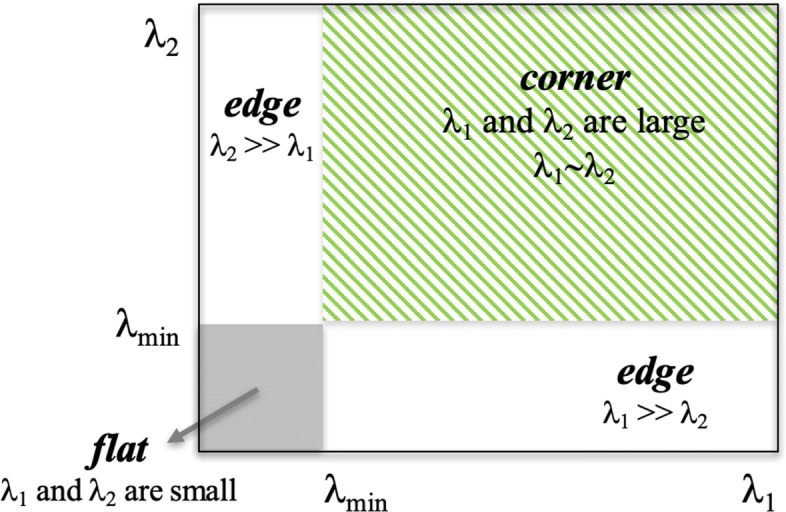
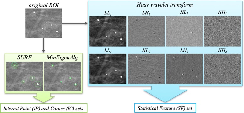
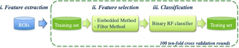
For each ROI, we extracted textural features on Haar wavelet decompositions and also interest points and corners detected by using Speeded Up Robust Feature (SURF) and Minimum Eigenvalue Algorithm (MinEigenAlg). Then a Random Forest binary classifier is trained on a subset of a sub-set features selected by two different kinds of feature selection techniques, such as filter and embedded methods. We tested the proposed model on 260 ROIs extracted from digital mammograms of the BCDR public database. The best prediction performance for the normal/abnormal and benign/malignant problems reaches a median AUC value of 98.16% and 92.08%, and an accuracy of 97.31% and 88.46%, respectively. The experimental result was comparable with related work performance.

CONCLUSIONS

The best performing result obtained with embedded method is more parsimonious than the filter one. The SURF and MinEigen algorithms provide a strong informative content useful for the characterization of microcalcification clusters.

摘要

背景

筛查项目使用乳房 X 光摄影术作为早期发现乳腺癌的主要诊断工具。然而,对于放射科医生来说,某些病变(如微钙化)的诊断仍然具有挑战性。在本文中,我们提出了一种用于区分数字乳房 X 光片中组织的自动二分类模型,作为放射科医生的辅助工具。特别是,我们比较了不同方法在特征选择过程中对学习性能和所选特征的贡献。

结果

对于每个感兴趣区域(ROI),我们在 Haar 小波分解上提取纹理特征,还使用加速稳健特征(SURF)和最小特征值算法(MinEigenAlg)检测感兴趣点和角点。然后,随机森林二分类器在通过两种不同的特征选择技术(如过滤法和嵌入式方法)选择的子集中的特征子集上进行训练。我们在 BCDR 公共数据库的数字乳房 X 光片中提取的 260 个 ROI 上测试了所提出的模型。正常/异常和良性/恶性问题的最佳预测性能达到了中位数 AUC 值 98.16%和 92.08%,准确性分别为 97.31%和 88.46%。实验结果与相关工作的性能相当。

结论

嵌入式方法获得的最佳性能结果比过滤法更简洁。SURF 和 MinEigen 算法提供了对微钙化簇特征描述非常有用的丰富信息内容。

https://cdn.ncbi.nlm.nih.gov/pmc/blobs/8090/7069158/b202474cdb6c/12859_2020_3358_Fig6_HTML.jpg
https://cdn.ncbi.nlm.nih.gov/pmc/blobs/8090/7069158/9bb458452adc/12859_2020_3358_Fig1_HTML.jpg
https://cdn.ncbi.nlm.nih.gov/pmc/blobs/8090/7069158/90c9c1cb7358/12859_2020_3358_Fig2_HTML.jpg
https://cdn.ncbi.nlm.nih.gov/pmc/blobs/8090/7069158/8e37e53d21f7/12859_2020_3358_Fig3_HTML.jpg
https://cdn.ncbi.nlm.nih.gov/pmc/blobs/8090/7069158/10e76ea3dcdf/12859_2020_3358_Fig4_HTML.jpg
https://cdn.ncbi.nlm.nih.gov/pmc/blobs/8090/7069158/2686e5a31eed/12859_2020_3358_Fig5_HTML.jpg
https://cdn.ncbi.nlm.nih.gov/pmc/blobs/8090/7069158/b202474cdb6c/12859_2020_3358_Fig6_HTML.jpg

相似文献

1
A machine learning approach on multiscale texture analysis for breast microcalcification diagnosis.基于多尺度纹理分析的机器学习方法在乳腺微钙化诊断中的应用。
BMC Bioinformatics. 2020 Mar 11;21(Suppl 2):91. doi: 10.1186/s12859-020-3358-4.
2
Breast microcalcifications detection based on fusing features with DTCWT.基于 DTCWT 融合特征的乳腺微钙化检测。
J Xray Sci Technol. 2020;28(2):197-218. doi: 10.3233/XST-190583.
3
Breast cancer diagnosis: analyzing texture of tissue surrounding microcalcifications.乳腺癌诊断:分析微钙化周围组织的纹理
IEEE Trans Inf Technol Biomed. 2008 Nov;12(6):731-8. doi: 10.1109/TITB.2008.920634.
4
A swarm optimized neural network system for classification of microcalcification in mammograms.基于群智能优化神经网络的乳腺钼靶微钙化分类系统。
J Med Syst. 2012 Oct;36(5):3051-61. doi: 10.1007/s10916-011-9781-3. Epub 2011 Sep 23.
5
A novel approach for detection and classification of mammographic microcalcifications using wavelet analysis and extreme learning machine.基于小波分析和极限学习机的乳腺微钙化检测与分类新方法。
Comput Biol Med. 2012 Sep;42(9):898-905. doi: 10.1016/j.compbiomed.2012.07.001. Epub 2012 Aug 4.
6
A novel machine learning approach on texture analysis for automatic breast microcalcification diagnosis classification of mammogram images.一种基于纹理分析的新型机器学习方法,用于自动乳腺微钙化诊断分类的乳腺 X 线图像。
J Cancer Res Clin Oncol. 2023 Aug;149(9):6151-6170. doi: 10.1007/s00432-023-04571-y. Epub 2023 Jan 21.
7
Computerized analysis of mammographic microcalcifications in morphological and texture feature spaces.乳腺钼靶微钙化在形态学和纹理特征空间中的计算机分析
Med Phys. 1998 Oct;25(10):2007-19. doi: 10.1118/1.598389.
8
SVM based system for classification of microcalcifications in digital mammograms.基于支持向量机的数字乳腺X线摄影中微钙化分类系统。
Conf Proc IEEE Eng Med Biol Soc. 2006;2006:4747-50. doi: 10.1109/IEMBS.2006.259320.
9
Mass detection in digital breast tomosynthesis: Deep convolutional neural network with transfer learning from mammography.数字乳腺断层合成中的肿块检测:基于乳腺X线摄影迁移学习的深度卷积神经网络
Med Phys. 2016 Dec;43(12):6654. doi: 10.1118/1.4967345.
10
Quantitative comparison of clustered microcalcifications in for-presentation and for-processing mammograms in full-field digital mammography.全数字化乳腺摄影中用于诊断和存档的钼靶片中小钙化簇的定量比较。
Med Phys. 2017 Jul;44(7):3726-3738. doi: 10.1002/mp.12316. Epub 2017 Jun 9.

引用本文的文献

1
Interpretable Radiomic Signature for Breast Microcalcification Detection and Classification.可解释的乳腺微钙化检测和分类放射组学特征。
J Imaging Inform Med. 2024 Jun;37(3):1038-1053. doi: 10.1007/s10278-024-01012-1. Epub 2024 Feb 13.
2
The application of traditional machine learning and deep learning techniques in mammography: a review.传统机器学习和深度学习技术在乳腺钼靶摄影中的应用:综述
Front Oncol. 2023 Aug 11;13:1213045. doi: 10.3389/fonc.2023.1213045. eCollection 2023.
3
A novel machine learning approach on texture analysis for automatic breast microcalcification diagnosis classification of mammogram images.

本文引用的文献

1
Global cancer statistics 2018: GLOBOCAN estimates of incidence and mortality worldwide for 36 cancers in 185 countries.全球癌症统计数据 2018:GLOBOCAN 对全球 185 个国家/地区 36 种癌症的发病率和死亡率的估计。
CA Cancer J Clin. 2018 Nov;68(6):394-424. doi: 10.3322/caac.21492. Epub 2018 Sep 12.
2
Annual Report to the Nation on the Status of Cancer, part I: National cancer statistics.《国家癌症报告:第一部分:全国癌症统计数据》
Cancer. 2018 Jul 1;124(13):2785-2800. doi: 10.1002/cncr.31551. Epub 2018 May 22.
3
Topological modeling and classification of mammographic microcalcification clusters.
一种基于纹理分析的新型机器学习方法,用于自动乳腺微钙化诊断分类的乳腺 X 线图像。
J Cancer Res Clin Oncol. 2023 Aug;149(9):6151-6170. doi: 10.1007/s00432-023-04571-y. Epub 2023 Jan 21.
4
Contrast-Enhanced Mammography (CEM) Capability to Distinguish Molecular Breast Cancer Subtypes.对比增强乳腺钼靶摄影(CEM)区分分子乳腺癌亚型的能力。
Biomedicines. 2022 Sep 24;10(10):2384. doi: 10.3390/biomedicines10102384.
5
Transfer learning approach based on computed tomography images for predicting late xerostomia after radiotherapy in patients with oropharyngeal cancer.基于计算机断层扫描图像的迁移学习方法用于预测口咽癌患者放疗后的晚期口干症。
Front Med (Lausanne). 2022 Sep 23;9:993395. doi: 10.3389/fmed.2022.993395. eCollection 2022.
6
A New Collaborative Classification Process for Microcalcification Detection Based on Graphs and Knowledge Propagation.基于图和知识传播的微钙化检测新协同分类过程。
J Digit Imaging. 2022 Dec;35(6):1560-1575. doi: 10.1007/s10278-022-00678-9. Epub 2022 Aug 1.
7
Diagnostic Strategies for Breast Cancer Detection: From Image Generation to Classification Strategies Using Artificial Intelligence Algorithms.乳腺癌检测的诊断策略:从图像生成到使用人工智能算法的分类策略
Cancers (Basel). 2022 Jul 15;14(14):3442. doi: 10.3390/cancers14143442.
8
Classification of Mammographic ROI for Microcalcification Detection Using Multifractal Approach.基于多重分形方法的乳腺 ROI 内微钙化检测分类。
J Digit Imaging. 2022 Dec;35(6):1544-1559. doi: 10.1007/s10278-022-00677-w. Epub 2022 Jul 19.
9
Automated quantitative assessment of amorphous calcifications: Towards improved malignancy risk stratification.自动化定量评估非晶形钙化:提高恶性风险分层。
Comput Biol Med. 2022 Jul;146:105504. doi: 10.1016/j.compbiomed.2022.105504. Epub 2022 Apr 8.
10
Digital subtraction of temporally sequential mammograms for improved detection and classification of microcalcifications.数字化减影时间序列乳腺 X 线摄影在微钙化检测和分类中的应用。
Eur Radiol Exp. 2021 Sep 14;5(1):40. doi: 10.1186/s41747-021-00238-w.
乳腺钼靶微钙化簇的拓扑建模与分类
IEEE Trans Biomed Eng. 2015 Apr;62(4):1203-14. doi: 10.1109/TBME.2014.2385102.
4
Wavelet-based 3D reconstruction of microcalcification clusters from two mammographic views: new evidence that fractal tumors are malignant and Euclidean tumors are benign.基于小波的从两个乳腺X线视图对微钙化簇进行三维重建:分形肿瘤为恶性而欧几里得肿瘤为良性的新证据。
PLoS One. 2014 Sep 15;9(9):e107580. doi: 10.1371/journal.pone.0107580. eCollection 2014.
5
Computer aided detection system for micro calcifications in digital mammograms.数字乳腺钼靶片中微钙化的计算机辅助检测系统
Comput Methods Programs Biomed. 2014 Oct;116(3):226-35. doi: 10.1016/j.cmpb.2014.04.010. Epub 2014 Apr 30.
6
Mammographic images segmentation based on chaotic map clustering algorithm.基于混沌图聚类算法的乳腺 X 光图像分割。
BMC Med Imaging. 2014 Mar 25;14:12. doi: 10.1186/1471-2342-14-12.
7
Computer-aided diagnosis of breast microcalcifications based on dual-tree complex wavelet transform.基于双树复小波变换的乳腺微钙化计算机辅助诊断。
Biomed Eng Online. 2012 Dec 19;11:96. doi: 10.1186/1475-925X-11-96.
8
Computer-aided detection/diagnosis of breast cancer in mammography and ultrasound: a review.计算机辅助检测/诊断乳腺癌在乳腺 X 线摄影和超声中的应用:综述。
Clin Imaging. 2013 May-Jun;37(3):420-6. doi: 10.1016/j.clinimag.2012.09.024. Epub 2012 Nov 13.
9
A novel approach for detection and classification of mammographic microcalcifications using wavelet analysis and extreme learning machine.基于小波分析和极限学习机的乳腺微钙化检测与分类新方法。
Comput Biol Med. 2012 Sep;42(9):898-905. doi: 10.1016/j.compbiomed.2012.07.001. Epub 2012 Aug 4.
10
Is single reading with computer-aided detection (CAD) as good as double reading in mammography screening? A systematic review.单读与计算机辅助检测 (CAD) 在乳腺筛查中的效果是否与双读相当?一项系统评价。
BMC Med Imaging. 2012 Jul 24;12:22. doi: 10.1186/1471-2342-12-22.