Cardiovascular Research Group, School of Human Sciences (Exercise and Sport Science), University of Western Australia, Perth, Australia.
Australian Research Council Centre of Excellence for Nanoscale Biophotonics, School of Biomedicine, Faculty of Health and Medical Sciences, University of Adelaide, Adelaide, Australia.
J Physiol. 2022 Sep;600(17):3921-3929. doi: 10.1113/JP282940. Epub 2022 Aug 10.
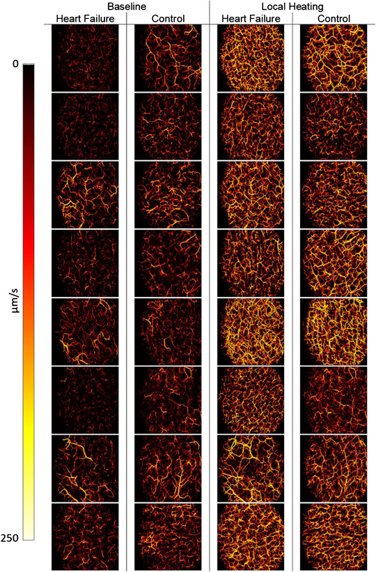
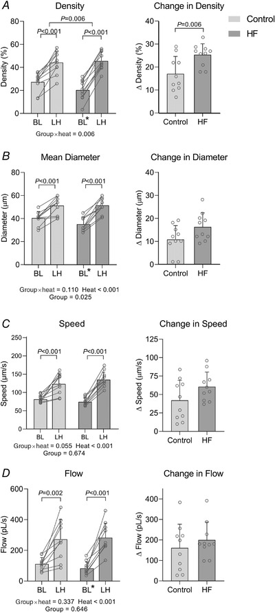
Heart failure (HF) is characterised by abnormal conduit and resistance artery function in humans. Microvascular function in HF is less well characterised, due in part to the lack of tools to image these vessels in vivo. The skin microvasculature is a surrogate for systemic microvascular function and health and plays a key role in thermoregulation, which is dysfunctional in HF. We deployed a novel optical coherence tomography (OCT) technique to visualise and quantify microvascular structure and function in 10 subjects with HF and 10 age- and sex-matched controls. OCT images were obtained from the ventral aspect of the forearm, at baseline (33°C) and after 30 min of localised skin heating. At rest, OCT-derived microvascular density (20.3 ± 8.7%, P = 0.004), diameter (35.1 ± 6.0 μm, P = 0.006) and blood flow (82.9 ± 41.1 pl/s, P = 0.021) were significantly lower in HF than CON (27.2 ± 8.0%, 40.4 ± 5.8 μm, 110.8 ± 41.9 pl/s), whilst blood speed was not significantly lower (74.3 ± 11.0 μm/s vs. 81.3 ± 9.9 μm/s, P = 0.069). After local heating, the OCT-based density, diameter, blood speed and blood flow of HF patients were similar (all P > 0.05) to CON. Although abnormalities exist at rest which may reflect microvascular disease status, patients with HF retain the capacity to dilate cutaneous microvessels in response to localised heat stress. This is a novel in vivo human observation of microvascular dysfunction in HF, illustrating the feasibility of OCT to directly visualise and quantify microvascular responses to physiological stimuli in vivo. KEY POINTS: Microvessels in the skin are critical to human thermoregulation, which is compromised in participants with heart failure (HF). We have developed a powerful new non-invasive optical coherence tomography (OCT)-based approach for the study of microvascular structure and function in vivo. Our approach enabled us to observe and quantify abnormal resting microvascular function in participants with HF. Patients with HF were able to dilate skin microvessels in response to local heat stress, arguing against an underlying structural abnormality. This suggests that microvascular functional regulation is the primary abnormality in HF. OCT can be used to directly visualise and quantify microvascular responses to physiological stimuli in vivo.
心力衰竭(HF)的特征是人类的传导和阻力动脉功能异常。HF 中的微血管功能描述得较少,部分原因是缺乏在体内成像这些血管的工具。皮肤微血管是全身微血管功能和健康的替代物,在体温调节中起着关键作用,而体温调节在 HF 中是功能失调的。我们部署了一种新的光学相干断层扫描(OCT)技术,从 10 名 HF 患者和 10 名年龄和性别匹配的对照者的前臂腹侧获得基线(33°C)和局部皮肤加热 30 分钟后的微血管结构和功能的 OCT 图像。在休息时,HF 中的 OCT 衍生微血管密度(20.3±8.7%,P=0.004)、直径(35.1±6.0μm,P=0.006)和血流(82.9±41.1pl/s,P=0.021)明显低于 CON(27.2±8.0%,40.4±5.8μm,110.8±41.9pl/s),而血流速度没有明显降低(74.3±11.0μm/s 与 81.3±9.9μm/s,P=0.069)。局部加热后,HF 患者的 OCT 基于的密度、直径、血流速度和血流与 CON 相似(均 P>0.05)。尽管在休息时存在异常,这可能反映了微血管疾病状态,但 HF 患者保留了对局部热应激扩张皮肤微血管的能力。这是 HF 中微血管功能障碍的一种新的体内人类观察,说明了 OCT 直接可视化和量化体内生理刺激下微血管反应的可行性。要点:皮肤中的微血管对人类体温调节至关重要,而 HF 患者的体温调节受损。我们开发了一种强大的新的非侵入性光学相干断层扫描(OCT)方法,用于研究体内微血管结构和功能。我们的方法使我们能够观察和量化 HF 患者静息时异常的微血管功能。HF 患者能够对局部热应激扩张皮肤微血管,这表明不存在潜在的结构异常。这表明微血管功能调节是 HF 的主要异常。OCT 可用于直接可视化和量化体内生理刺激下的微血管反应。