Heart Group, Faculty of Medicine and Health Technology, Tampere University, Tampere, Finland.
Biomaterials and Tissue Engineering Group, Faculty of Medicine and Health Technology, Tampere University, Tampere, Finland.
Cell Biol Toxicol. 2023 Feb;39(1):145-163. doi: 10.1007/s10565-022-09742-0. Epub 2022 Jul 23.
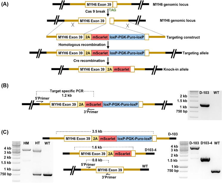
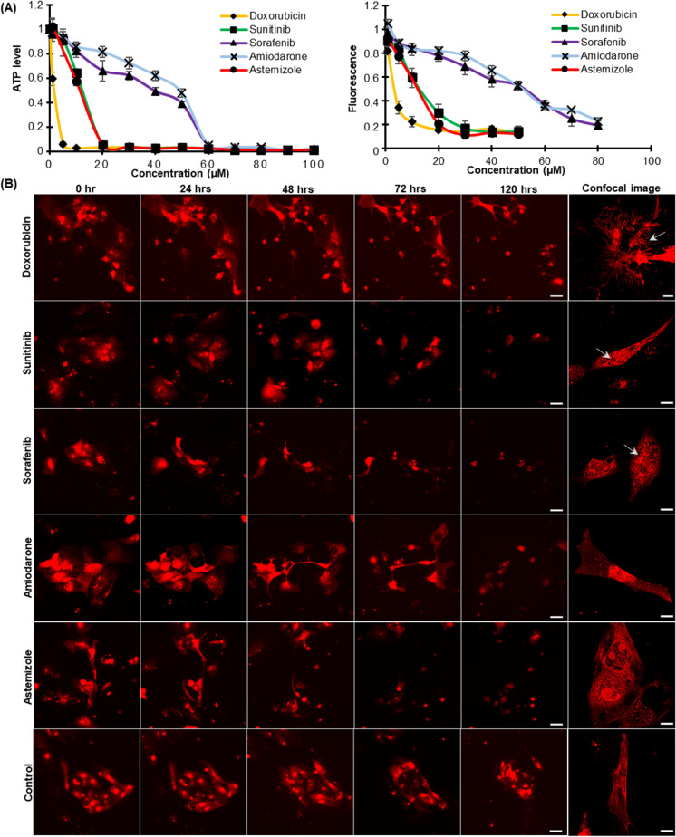
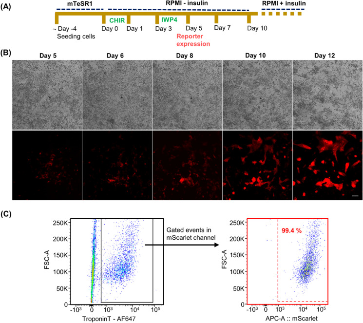
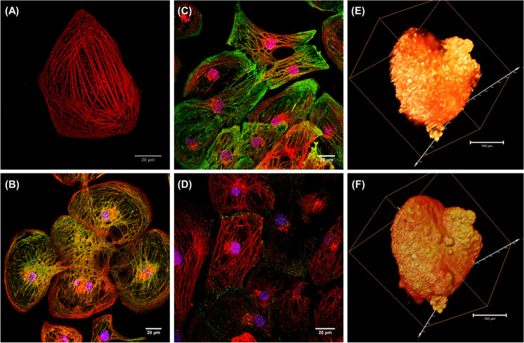
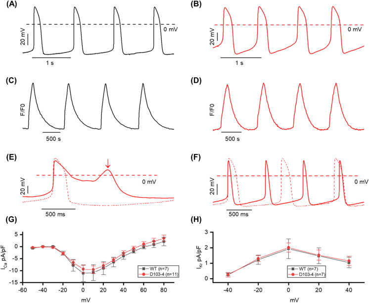
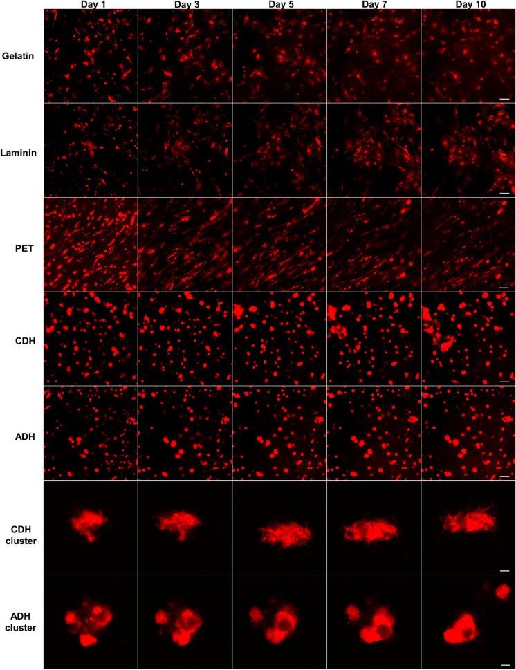
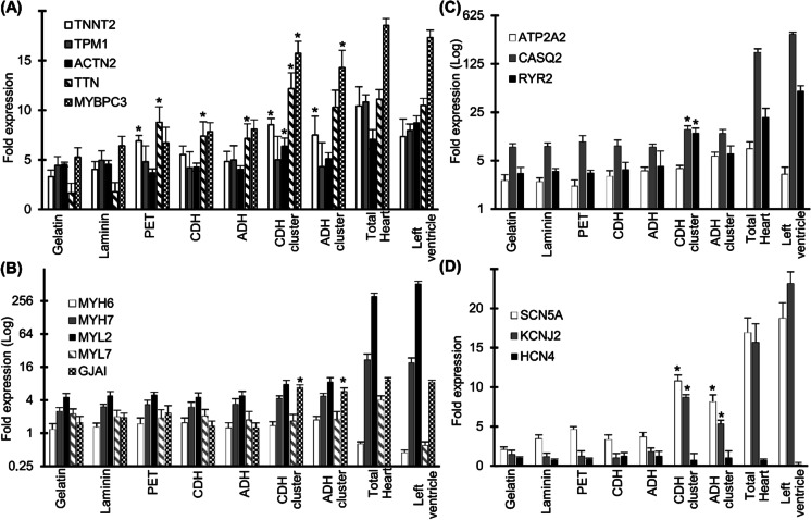
Human induced pluripotent stem cell derived cardiomyocytes (hiPSC-CMs) hold great potential in the cardiovascular field for human disease modeling, drug development, and regenerative medicine. However, multiple hurdles still exist for the effective utilization of hiPSC-CMs as a human-based experimental platform that can be an alternative to the current animal models. To further expand their potential as a research tool and bridge the translational gap, we have generated a cardiac-specific hiPSC reporter line that differentiates into fluorescent CMs using CRISPR-Cas9 genome editing technology. The CMs illuminated with the mScarlet fluorescence enable their non-invasive continuous tracking and functional cellular phenotyping, offering a real-time 2D/3D imaging platform. Utilizing the reporter CMs, we developed an imaging-based cardiotoxicity screening system that can monitor distinct drug-induced structural toxicity and CM viability in real time. The reporter fluorescence enabled visualization of sarcomeric disarray and displayed a drug dose-dependent decrease in its fluorescence. The study also has demonstrated the reporter CMs as a biomaterial cytocompatibility analysis tool that can monitor dynamic cell behavior and maturity of hiPSC-CMs cultured in various biomaterial scaffolds. This versatile cardiac imaging tool that enables real time tracking and high-resolution imaging of CMs has significant potential in disease modeling, drug screening, and toxicology testing.
人诱导多能干细胞衍生的心肌细胞(hiPSC-CMs)在心血管领域具有巨大的潜力,可用于人类疾病建模、药物开发和再生医学。然而,要将 hiPSC-CMs 有效用作替代当前动物模型的基于人类的实验平台,仍然存在多个障碍。为了进一步扩大其作为研究工具的潜力并缩小转化差距,我们使用 CRISPR-Cas9 基因组编辑技术生成了一种心脏特异性 hiPSC 报告细胞系,该细胞系可分化为荧光心肌细胞。用 mScarlet 荧光标记的心肌细胞可实现其非侵入性的连续跟踪和功能细胞表型分析,提供了一个实时的 2D/3D 成像平台。利用报告细胞,我们开发了一种基于成像的心脏毒性筛选系统,可实时监测不同药物引起的结构毒性和心肌细胞活力。报告荧光可使肌节排列紊乱可视化,并显示其荧光随药物剂量的依赖性降低。该研究还表明,报告细胞可作为生物材料细胞相容性分析工具,用于监测在各种生物材料支架中培养的 hiPSC-CMs 的动态细胞行为和成熟度。这种多功能的心脏成像工具能够实时跟踪和高分辨率成像心肌细胞,在疾病建模、药物筛选和毒理学测试方面具有重要的应用潜力。