Lundberg Laboratory for Diabetes Research, Department of Molecular and Clinical Medicine, Sahlgrenska Academy, University of Gothenburg, Gothenburg, Sweden.
Lundberg Laboratory for Diabetes Research, Department of Molecular and Clinical Medicine, Sahlgrenska Academy, University of Gothenburg, Gothenburg, Sweden; Department of Translational Medical Sciences, Federico II University of Naples, Naples, Italy; URT Genomics of Diabetes, Institute of Experimental Endocrinology and Oncology, National Research Council, Naples, Italy.
Mol Metab. 2022 Oct;64:101558. doi: 10.1016/j.molmet.2022.101558. Epub 2022 Jul 21.
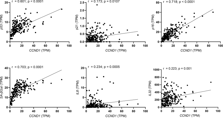
Cellular senescence, an irreversible proliferative cell arrest, is caused by excessive intracellular or extracellular stress/damage. Increased senescent cells have been identified in multiple tissues in different metabolic and other aging-related diseases. Recently, several human and mouse studies emphasized the involvement of senescence in development and progression of NAFLD. Hyperinsulinemia, seen in obesity, metabolic syndrome, and other conditions of insulin resistance, has been linked to senescence in adipocytes and neurons. Here, we investigate the possible direct role of chronic hyperinsulinemia in the development of senescence in human hepatocytes.
Using fluorescence microscopy, immunoblotting, and gene expression, we tested senescence markers in human hepatocytes subjected to chronic hyperinsulinemia in vitro and validated the data in vivo by using liver-specific insulin receptor knockout (LIRKO) mice. The consequences of hyperinsulinemia were also studied in senescent hepatocytes following doxorubicin as a model of stress-induced senescence. Furthermore, the effects of senolytic agents in insulin- and doxorubicin-treated cells were analyzed.
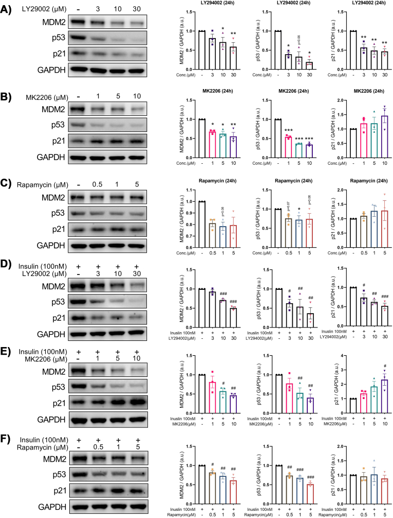
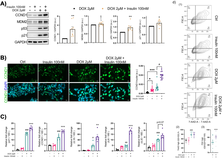
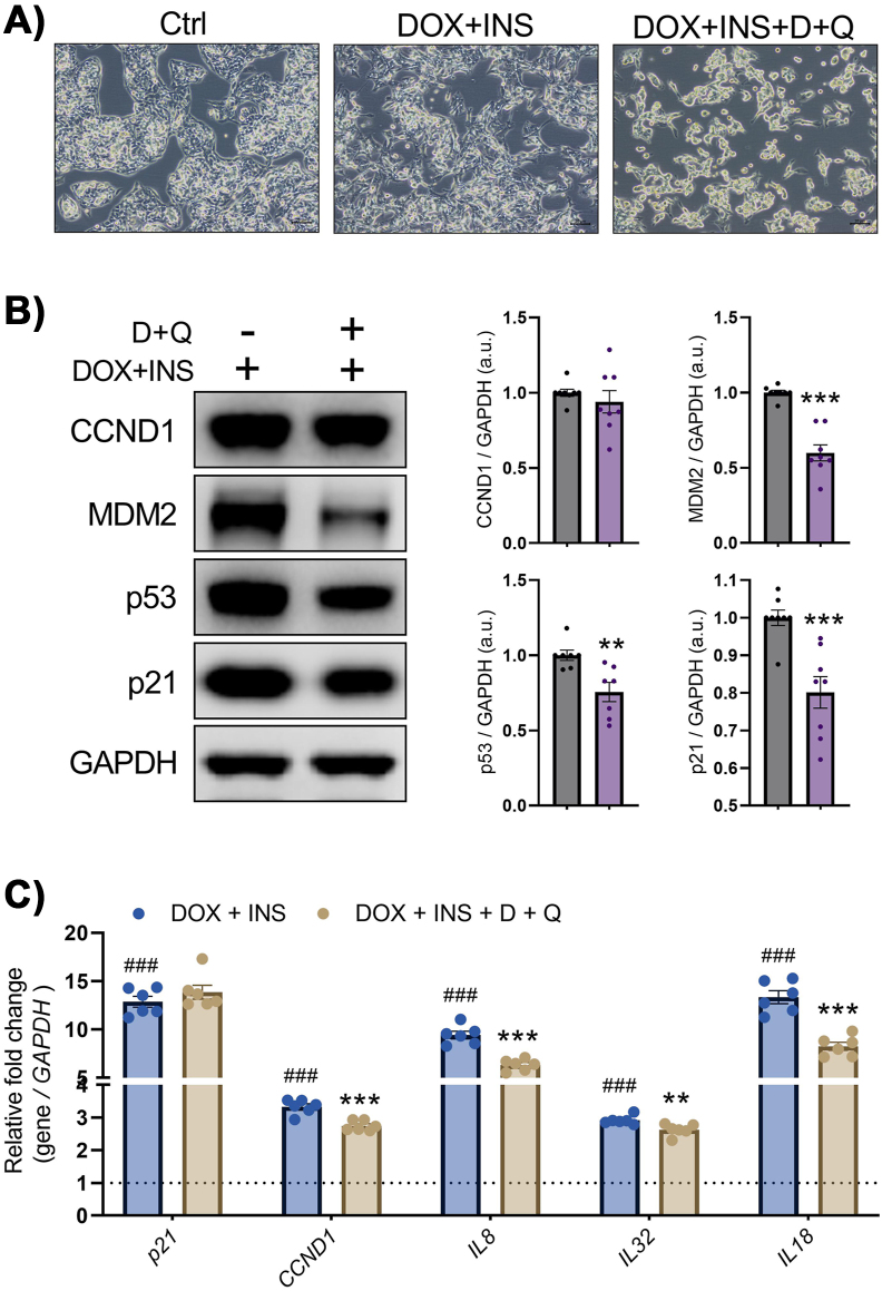
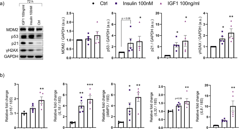
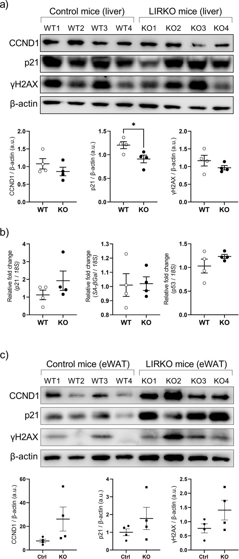
Results showed that exposing the hepatocytes to prolonged hyperinsulinemia promotes the onset of senescence by increasing the expression of p53 and p21. It also further enhanced the senescent phenotype in already senescent hepatocytes. Addition of insulin signaling pathway inhibitors prevented the increase in cell senescence, supporting the direct contribution of insulin. Furthermore, LIRKO mice, in which insulin signaling in the liver is abolished due to deletion of the insulin receptor gene, showed no differences in senescence compared to their wild-type counterparts despite having marked hyperinsulinemia indicating these are receptor-mediated effects. In contrast, the persistent hyperinsulinemia in LIRKO mice enhanced senescence in white adipose tissue. In vitro, senolytic agents dasatinib and quercetin reduced the prosenescent effects of hyperinsulinemia in hepatocytes.
Our findings demonstrate a direct link between chronic hyperinsulinemia and hepatocyte senescence. This effect can be blocked by reducing the levels of insulin receptors or administration of senolytic drugs, such as dasatinib and quercetin.
细胞衰老,即细胞不可逆的增殖停滞,是由细胞内或细胞外过度的应激/损伤引起的。在多种组织中,在不同的代谢和其他与衰老相关的疾病中,已经发现了更多的衰老细胞。最近,几项人类和小鼠研究强调了衰老在非酒精性脂肪性肝病(NAFLD)的发展和进展中的作用。肥胖症、代谢综合征和其他胰岛素抵抗情况下的高胰岛素血症,与脂肪细胞和神经元中的衰老有关。在这里,我们研究了慢性高胰岛素血症在人类肝细胞衰老发展中的可能直接作用。
我们通过荧光显微镜、免疫印迹和基因表达,检测了体外慢性高胰岛素血症作用下的人肝细胞中的衰老标志物,并通过使用肝特异性胰岛素受体敲除(LIRKO)小鼠在体内验证了这些数据。我们还研究了多柔比星作为应激诱导衰老模型作用下衰老肝细胞中高胰岛素血症的后果。此外,还分析了衰老细胞中胰岛素和多柔比星处理后的细胞对衰老溶解剂的反应。
结果表明,长期暴露于高胰岛素血症会通过增加 p53 和 p21 的表达来促进衰老的发生。它还进一步增强了已经衰老的肝细胞中的衰老表型。胰岛素信号通路抑制剂的添加阻止了细胞衰老的增加,支持了胰岛素的直接作用。此外,由于胰岛素受体基因缺失而导致肝脏中胰岛素信号被阻断的 LIRKO 小鼠与野生型小鼠相比,衰老没有差异,尽管它们的胰岛素水平明显升高,这表明这些是受体介导的效应。相比之下,LIRKO 小鼠中的持续高胰岛素血症增强了白色脂肪组织中的衰老。在体外,衰老溶解剂达沙替尼和槲皮素减少了高胰岛素血症对肝细胞的促衰老作用。
我们的研究结果表明,慢性高胰岛素血症与肝细胞衰老之间存在直接联系。这种作用可以通过降低胰岛素受体水平或使用达沙替尼和槲皮素等衰老溶解药物来阻断。