• 文献检索
  • 文档翻译
  • 深度研究
  • 学术资讯
  • Suppr Zotero 插件Zotero 插件
  • 邀请有礼
  • 套餐&价格
  • 历史记录
应用&插件
Suppr Zotero 插件Zotero 插件浏览器插件Mac 客户端Windows 客户端微信小程序
定价
高级版会员购买积分包购买API积分包
服务
文献检索文档翻译深度研究API 文档MCP 服务
关于我们
关于 Suppr公司介绍联系我们用户协议隐私条款
关注我们

Suppr 超能文献

核心技术专利:CN118964589B侵权必究
粤ICP备2023148730 号-1Suppr @ 2026

文献检索

告别复杂PubMed语法,用中文像聊天一样搜索,搜遍4000万医学文献。AI智能推荐,让科研检索更轻松。

立即免费搜索

文件翻译

保留排版,准确专业,支持PDF/Word/PPT等文件格式,支持 12+语言互译。

免费翻译文档

深度研究

AI帮你快速写综述,25分钟生成高质量综述,智能提取关键信息,辅助科研写作。

立即免费体验

StainCUT:基于对比学习的染色归一化

StainCUT: Stain Normalization with Contrastive Learning.

作者信息

Gutiérrez Pérez José Carlos, Otero Baguer Daniel, Maass Peter

机构信息

Center for Industrial Mathematics, University of Bremen, 28359 Bremen, Germany.

出版信息

J Imaging. 2022 Jul 20;8(7):202. doi: 10.3390/jimaging8070202.

DOI:10.3390/jimaging8070202
PMID:35877646
原文链接:https://pmc.ncbi.nlm.nih.gov/articles/PMC9317097/
Abstract

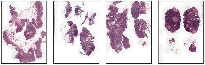
In recent years, numerous deep-learning approaches have been developed for the analysis of histopathology Whole Slide Images (WSI). A recurrent issue is the lack of generalization ability of a model that has been trained with images of one laboratory and then used to analyze images of a different laboratory. This occurs mainly due to the use of different scanners, laboratory procedures, and staining variations. This can produce strong color differences, which change not only the characteristics of the image, such as the contrast, brightness, and saturation, but also create more complex style variations. In this paper, we present a deep-learning solution based on contrastive learning to transfer from one staining style to another: StainCUT. This method eliminates the need to choose a reference frame and does not need paired images with different staining to learn the mapping between the stain distributions. Additionally, it does not rely on the CycleGAN approach, which makes the method efficient in terms of memory consumption and running time. We evaluate the model using two datasets that consist of the same specimens digitized with two different scanners. We also apply it as a preprocessing step for the semantic segmentation of metastases in lymph nodes. The model was trained on data from one of the laboratories and evaluated on data from another. The results validate the hypothesis that stain normalization indeed improves the performance of the model. Finally, we also investigate and compare the application of the stain normalization step during the training of the model and at inference.

摘要

近年来,已经开发了许多深度学习方法用于组织病理学全切片图像(WSI)的分析。一个反复出现的问题是,一个在一个实验室的图像上训练,然后用于分析另一个实验室图像的模型缺乏泛化能力。这种情况主要是由于使用了不同的扫描仪、实验室程序和染色差异。这会产生强烈的颜色差异,不仅会改变图像的特征,如对比度、亮度和饱和度,还会产生更复杂的风格变化。在本文中,我们提出了一种基于对比学习的深度学习解决方案,用于从一种染色风格转换到另一种染色风格:StainCUT。该方法无需选择参考帧,也不需要具有不同染色的配对图像来学习染色分布之间的映射。此外,它不依赖于CycleGAN方法,这使得该方法在内存消耗和运行时间方面都很高效。我们使用两个数据集对模型进行评估,这两个数据集由用两种不同扫描仪数字化的相同标本组成。我们还将其作为淋巴结转移语义分割的预处理步骤应用。该模型在其中一个实验室的数据上进行训练,并在另一个实验室的数据上进行评估。结果验证了染色归一化确实能提高模型性能的假设。最后,我们还研究并比较了染色归一化步骤在模型训练期间和推理时的应用。

https://cdn.ncbi.nlm.nih.gov/pmc/blobs/0d2e/9317097/688c4a644a44/jimaging-08-00202-g015.jpg
https://cdn.ncbi.nlm.nih.gov/pmc/blobs/0d2e/9317097/7a99980919cb/jimaging-08-00202-g0A1.jpg
https://cdn.ncbi.nlm.nih.gov/pmc/blobs/0d2e/9317097/cae94ca03535/jimaging-08-00202-g0A2.jpg
https://cdn.ncbi.nlm.nih.gov/pmc/blobs/0d2e/9317097/c10ec21db986/jimaging-08-00202-g001.jpg
https://cdn.ncbi.nlm.nih.gov/pmc/blobs/0d2e/9317097/7de7e6b40c48/jimaging-08-00202-g002.jpg
https://cdn.ncbi.nlm.nih.gov/pmc/blobs/0d2e/9317097/ed81d053f1b8/jimaging-08-00202-g003.jpg
https://cdn.ncbi.nlm.nih.gov/pmc/blobs/0d2e/9317097/2394df9fe2a0/jimaging-08-00202-g004.jpg
https://cdn.ncbi.nlm.nih.gov/pmc/blobs/0d2e/9317097/44a02540fb5f/jimaging-08-00202-g005.jpg
https://cdn.ncbi.nlm.nih.gov/pmc/blobs/0d2e/9317097/42544709d932/jimaging-08-00202-g006.jpg
https://cdn.ncbi.nlm.nih.gov/pmc/blobs/0d2e/9317097/2bf10bb8f9aa/jimaging-08-00202-g007.jpg
https://cdn.ncbi.nlm.nih.gov/pmc/blobs/0d2e/9317097/4c5c5bfbbafb/jimaging-08-00202-g008.jpg
https://cdn.ncbi.nlm.nih.gov/pmc/blobs/0d2e/9317097/cccd9a9e5657/jimaging-08-00202-g009.jpg
https://cdn.ncbi.nlm.nih.gov/pmc/blobs/0d2e/9317097/41d2fd055506/jimaging-08-00202-g010.jpg
https://cdn.ncbi.nlm.nih.gov/pmc/blobs/0d2e/9317097/7952a75fa9b1/jimaging-08-00202-g011.jpg
https://cdn.ncbi.nlm.nih.gov/pmc/blobs/0d2e/9317097/4fba9d1f7b72/jimaging-08-00202-g012.jpg
https://cdn.ncbi.nlm.nih.gov/pmc/blobs/0d2e/9317097/ccf6b7ee9563/jimaging-08-00202-g013.jpg
https://cdn.ncbi.nlm.nih.gov/pmc/blobs/0d2e/9317097/47b2d0217da1/jimaging-08-00202-g014.jpg
https://cdn.ncbi.nlm.nih.gov/pmc/blobs/0d2e/9317097/688c4a644a44/jimaging-08-00202-g015.jpg

相似文献

1
StainCUT: Stain Normalization with Contrastive Learning.StainCUT:基于对比学习的染色归一化
J Imaging. 2022 Jul 20;8(7):202. doi: 10.3390/jimaging8070202.
2
Normalization of HE-stained histological images using cycle consistent generative adversarial networks.使用循环一致生成对抗网络对 HE 染色组织学图像进行归一化。
Diagn Pathol. 2021 Aug 6;16(1):71. doi: 10.1186/s13000-021-01126-y.
3
The role of unpaired image-to-image translation for stain color normalization in colorectal cancer histology classification.非配对图像到图像翻译在结直肠癌组织学分类中用于染色颜色归一化的作用。
Comput Methods Programs Biomed. 2023 Jun;234:107511. doi: 10.1016/j.cmpb.2023.107511. Epub 2023 Mar 26.
4
Efficient Staining-Invariant Nuclei Segmentation Approach Using Self-Supervised Deep Contrastive Network.基于自监督深度对比网络的高效染色不变细胞核分割方法
Diagnostics (Basel). 2022 Dec 2;12(12):3024. doi: 10.3390/diagnostics12123024.
5
Stain color translation of multi-domain OSCC histopathology images using attention gated cGAN.使用注意力门控条件生成对抗网络对多域口腔鳞状细胞癌组织病理学图像进行染色颜色转换
Comput Med Imaging Graph. 2023 Jun;106:102202. doi: 10.1016/j.compmedimag.2023.102202. Epub 2023 Feb 24.
6
StainNet: A Fast and Robust Stain Normalization Network.StainNet:一种快速且稳健的染色归一化网络。
Front Med (Lausanne). 2021 Nov 5;8:746307. doi: 10.3389/fmed.2021.746307. eCollection 2021.
7
Seamless Virtual Whole Slide Image Synthesis and Validation Using Perceptual Embedding Consistency.基于感知嵌入一致性的无缝虚拟全幻灯片图像合成与验证。
IEEE J Biomed Health Inform. 2021 Feb;25(2):403-411. doi: 10.1109/JBHI.2020.2975151. Epub 2021 Feb 5.
8
Data-driven color augmentation for H&E stained images in computational pathology.计算病理学中用于苏木精-伊红(H&E)染色图像的数据驱动颜色增强
J Pathol Inform. 2023 Jan 3;14:100183. doi: 10.1016/j.jpi.2022.100183. eCollection 2023.
9
Adversarial Stain Transfer for Histopathology Image Analysis.对抗性染色转移在组织病理学图像分析中的应用。
IEEE Trans Med Imaging. 2018 Mar;37(3):792-802. doi: 10.1109/TMI.2017.2781228.
10
Self-Attentive Adversarial Stain Normalization.自注意力对抗染色归一化
Pattern Recognit (2021). 2021 Jan;12661:120-140. doi: 10.1007/978-3-030-68763-2_10. Epub 2021 Feb 21.

引用本文的文献

1
Stain Normalization of Histopathological Images Based on Deep Learning: A Review.基于深度学习的组织病理学图像染色归一化:综述
Diagnostics (Basel). 2025 Apr 18;15(8):1032. doi: 10.3390/diagnostics15081032.
2
Deep learning-based virtual staining, segmentation, and classification in label-free photoacoustic histology of human specimens.基于深度学习的人体标本无标记光声组织学中的虚拟染色、分割和分类
Light Sci Appl. 2024 Sep 2;13(1):226. doi: 10.1038/s41377-024-01554-7.
3
Investigating Contrastive Pair Learning's Frontiers in Supervised, Semisupervised, and Self-Supervised Learning.

本文引用的文献

1
Deeply Supervised UNet for Semantic Segmentation to Assist Dermatopathological Assessment of Basal Cell Carcinoma.用于语义分割的深度监督UNet辅助基底细胞癌的皮肤病理学评估
J Imaging. 2021 Apr 13;7(4):71. doi: 10.3390/jimaging7040071.
2
Deep learning in histopathology: the path to the clinic.深度学习在组织病理学中的应用:通往临床的道路。
Nat Med. 2021 May;27(5):775-784. doi: 10.1038/s41591-021-01343-4. Epub 2021 May 14.
3
Assessing the Impact of Color Normalization in Convolutional Neural Network-Based Nuclei Segmentation Frameworks.
探究对比对学习在监督学习、半监督学习和自监督学习中的前沿进展。
J Imaging. 2024 Aug 13;10(8):196. doi: 10.3390/jimaging10080196.
评估颜色归一化在基于卷积神经网络的细胞核分割框架中的影响。
Front Bioeng Biotechnol. 2019 Nov 1;7:300. doi: 10.3389/fbioe.2019.00300. eCollection 2019.
4
Computational pathology definitions, best practices, and recommendations for regulatory guidance: a white paper from the Digital Pathology Association.计算病理学定义、最佳实践和监管指南建议:数字病理学协会白皮书。
J Pathol. 2019 Nov;249(3):286-294. doi: 10.1002/path.5331. Epub 2019 Sep 3.
5
1399 H&E-stained sentinel lymph node sections of breast cancer patients: the CAMELYON dataset.1399 例乳腺癌患者 H&E 染色前哨淋巴结切片:CAMELYON 数据集。
Gigascience. 2018 Jun 1;7(6). doi: 10.1093/gigascience/giy065.
6
Structure-Preserving Color Normalization and Sparse Stain Separation for Histological Images.结构保持的颜色归一化和组织学图像的稀疏染色分离。
IEEE Trans Med Imaging. 2016 Aug;35(8):1962-71. doi: 10.1109/TMI.2016.2529665. Epub 2016 Apr 27.
7
A method for normalizing pathology images to improve feature extraction for quantitative pathology.一种用于标准化病理图像以改善定量病理学特征提取的方法。
Med Phys. 2016 Jan;43(1):528. doi: 10.1118/1.4939130.
8
Stain Specific Standardization of Whole-Slide Histopathological Images.全切片组织病理学图像的染色特异性标准化
IEEE Trans Med Imaging. 2016 Feb;35(2):404-15. doi: 10.1109/TMI.2015.2476509. Epub 2015 Sep 4.
9
A nonlinear mapping approach to stain normalization in digital histopathology images using image-specific color deconvolution.一种使用特定图像颜色反卷积对数字组织病理学图像进行染色归一化的非线性映射方法。
IEEE Trans Biomed Eng. 2014 Jun;61(6):1729-38. doi: 10.1109/TBME.2014.2303294.
10
Image segmentation with implicit color standardization using spatially constrained expectation maximization: detection of nuclei.使用空间约束期望最大化进行隐式颜色标准化的图像分割:细胞核检测
Med Image Comput Comput Assist Interv. 2012;15(Pt 1):365-72. doi: 10.1007/978-3-642-33415-3_45.