• 文献检索
  • 文档翻译
  • 深度研究
  • 学术资讯
  • Suppr Zotero 插件Zotero 插件
  • 邀请有礼
  • 套餐&价格
  • 历史记录
应用&插件
Suppr Zotero 插件Zotero 插件浏览器插件Mac 客户端Windows 客户端微信小程序
定价
高级版会员购买积分包购买API积分包
服务
文献检索文档翻译深度研究API 文档MCP 服务
关于我们
关于 Suppr公司介绍联系我们用户协议隐私条款
关注我们

Suppr 超能文献

核心技术专利:CN118964589B侵权必究
粤ICP备2023148730 号-1Suppr @ 2026

文献检索

告别复杂PubMed语法,用中文像聊天一样搜索,搜遍4000万医学文献。AI智能推荐,让科研检索更轻松。

立即免费搜索

文件翻译

保留排版,准确专业,支持PDF/Word/PPT等文件格式,支持 12+语言互译。

免费翻译文档

深度研究

AI帮你快速写综述,25分钟生成高质量综述,智能提取关键信息,辅助科研写作。

立即免费体验

Ahnak 缺失通过调节 TGF-β/Smad 信号通路加速肝脏再生。

Ahnak depletion accelerates liver regeneration by modulating the TGF-β/Smad signaling pathway.

机构信息

Laboratory of Developmental Biology and Genomics, College of Veterinary Medicine, Seoul National University, Seoul 08826, Korea.

Department of Surgery, University of Michigan, Ann Arbor 48109, MI, USA.

出版信息

BMB Rep. 2022 Aug;55(8):401-406. doi: 10.5483/BMBRep.2022.55.8.071.

DOI:10.5483/BMBRep.2022.55.8.071
PMID:35880432
原文链接:https://pmc.ncbi.nlm.nih.gov/articles/PMC9442348/
Abstract

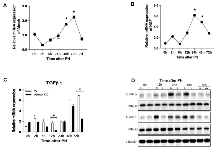
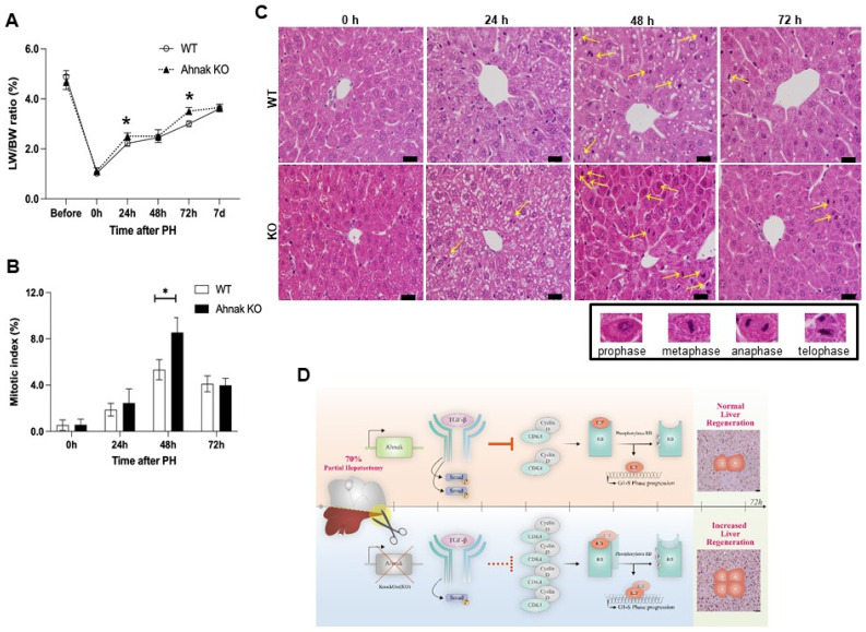
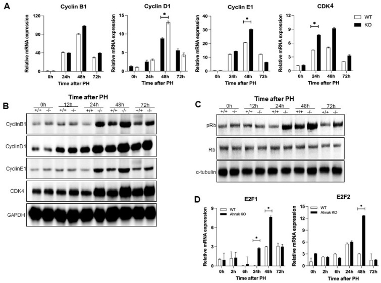
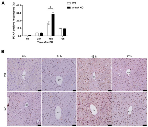
Ahnak, a large protein first identified as an inhibitor of TGF-β signaling in human neuroblastoma, was recently shown to promote TGF-β in some cancers. The TGF-β signaling pathway regulates cell growth, various biological functions, and cancer growth and metastasis. In this study, we used Ahnak knockout (KO) mice that underwent a 70% partial hepatectomy (PH) to investigate the function of Ahnak in TGF-β signaling during liver regeneration. At the indicated time points after PH, we analyzed the mRNA and protein expression of the TGF -β/Smad signaling pathway and cell cycle-related factors, evaluated the cell cycle through proliferating cell nuclear antigen (PCNA) immunostaining, analyzed the mitotic index by hematoxylin and eosin staining. We also measured the ratio of liver tissue weight to body weight. Activation of TGF-β signaling was confirmed by analyzing the levels of phospho-Smad 2 and 3 in the liver at the indicated time points after PH and was lower in Ahnak KO mice than in WT mice. The expression levels of cyclin B1, D1, and E1; proteins in the Rb/E2F transcriptional pathway, which regulates the cell cycle; and the numbers of PCNA-positive cells were increased in Ahnak KO mice and showed tendencies opposite that of TGF-β expression. During postoperative regeneration, the liver weight to body weight ratio tended to increase faster in Ahnak KO mice. However, 7 days after PH, both groups of mice showed similar rates of regeneration, following which their active regeneration stopped. Analysis of hepatocytes undergoing mitosis showed that there were more mitotic cells in Ahnak KO mice, consistent with the weight ratio. Our findings suggest that Ahnak enhances TGF-β signaling during postoperative liver regeneration, resulting in cell cycle disruption; this highlights a novel role of Ahnak in liver regeneration. These results provide new insight into liver regeneration and potential treatment targets for liver diseases that require surgical treatment. [BMB Reports 2022; 55(8): 401-406].

摘要

Ahnak,一种最初在人类神经母细胞瘤中被鉴定为 TGF-β 信号抑制剂的大型蛋白质,最近被证明在某些癌症中促进 TGF-β。TGF-β 信号通路调节细胞生长、各种生物学功能以及癌症的生长和转移。在这项研究中,我们使用接受 70%部分肝切除术(PH)的 Ahnak 敲除(KO)小鼠来研究 Ahnak 在 TGF-β 信号在肝再生过程中的功能。在 PH 后指定的时间点,我们分析了 TGF-β/Smad 信号通路和细胞周期相关因子的 mRNA 和蛋白表达,通过增殖细胞核抗原(PCNA)免疫染色评估细胞周期,通过苏木精和伊红染色分析有丝分裂指数。我们还测量了肝组织重量与体重的比值。通过分析 PH 后指定时间点肝中磷酸化 Smad2 和 3 的水平来确认 TGF-β 信号的激活,并且在 Ahnak KO 小鼠中比在 WT 小鼠中低。细胞周期调节转录途径中的细胞周期蛋白 B1、D1 和 E1 水平;Rb/E2F 转录途径中的蛋白;和 PCNA 阳性细胞的数量在 Ahnak KO 小鼠中增加,并表现出与 TGF-β 表达相反的趋势。在术后再生过程中,Ahnak KO 小鼠的肝重与体重比倾向于更快增加。然而,在 PH 后 7 天,两组小鼠的再生率相似,此后它们的活跃再生停止。对有丝分裂的肝细胞进行分析表明,Ahnak KO 小鼠中有更多的有丝分裂细胞,与体重比一致。我们的研究结果表明,Ahnak 在术后肝再生过程中增强 TGF-β 信号,导致细胞周期紊乱;这凸显了 Ahnak 在肝再生中的新作用。这些结果为肝再生提供了新的见解,并为需要手术治疗的肝脏疾病提供了潜在的治疗靶点。[BMB 报告 2022;55(8):401-406]。

https://cdn.ncbi.nlm.nih.gov/pmc/blobs/bea5/9442348/862b04a9dbc3/bmb-55-8-401-f4.jpg
https://cdn.ncbi.nlm.nih.gov/pmc/blobs/bea5/9442348/8d888805a693/bmb-55-8-401-f1.jpg
https://cdn.ncbi.nlm.nih.gov/pmc/blobs/bea5/9442348/5ea4e1274c29/bmb-55-8-401-f2.jpg
https://cdn.ncbi.nlm.nih.gov/pmc/blobs/bea5/9442348/f026881d2647/bmb-55-8-401-f3.jpg
https://cdn.ncbi.nlm.nih.gov/pmc/blobs/bea5/9442348/862b04a9dbc3/bmb-55-8-401-f4.jpg

相似文献

1
Ahnak depletion accelerates liver regeneration by modulating the TGF-β/Smad signaling pathway.Ahnak 缺失通过调节 TGF-β/Smad 信号通路加速肝脏再生。
BMB Rep. 2022 Aug;55(8):401-406. doi: 10.5483/BMBRep.2022.55.8.071.
2
Intact signaling by transforming growth factor beta is not required for termination of liver regeneration in mice.转化生长因子β的完整信号传导对于小鼠肝脏再生的终止并非必需。
Hepatology. 2004 Nov;40(5):1098-105. doi: 10.1002/hep.20426.
3
Inactivation of TGF-beta signaling in hepatocytes results in an increased proliferative response after partial hepatectomy.肝细胞中转化生长因子-β信号的失活导致部分肝切除术后增殖反应增强。
Oncogene. 2005 Apr 21;24(18):3028-41. doi: 10.1038/sj.onc.1208475.
4
Transforming growth factor-beta1 released from the spleen exerts a growth inhibitory effect on liver regeneration in rats.脾脏释放的转化生长因子β1对大鼠肝脏再生具有生长抑制作用。
Lab Invest. 2003 Nov;83(11):1595-603. doi: 10.1097/01.lab.0000095686.10639.c8.
5
Reticulon 4B (Nogo-B) facilitates hepatocyte proliferation and liver regeneration in mice.Reticulon 4B (Nogo-B) 促进小鼠肝细胞增殖和肝再生。
Hepatology. 2013 May;57(5):1992-2003. doi: 10.1002/hep.26235. Epub 2013 Mar 19.
6
Vitamin D3 up-regulated protein 1 deficiency accelerates liver regeneration after partial hepatectomy in mice.维生素 D3 上调蛋白 1 缺乏加速小鼠肝部分切除术后的肝再生。
J Hepatol. 2011 Jun;54(6):1168-76. doi: 10.1016/j.jhep.2010.09.025. Epub 2010 Oct 26.
7
Up-regulated transcriptional repressors SnoN and Ski bind Smad proteins to antagonize transforming growth factor-beta signals during liver regeneration.上调的转录抑制因子SnoN和Ski在肝脏再生过程中与Smad蛋白结合,拮抗转化生长因子-β信号。
J Biol Chem. 2002 Aug 9;277(32):28483-90. doi: 10.1074/jbc.M202403200. Epub 2002 May 21.
8
Ahnak functions as a tumor suppressor via modulation of TGFβ/Smad signaling pathway.Ahnak通过调节TGFβ/Smad信号通路发挥肿瘤抑制作用。
Oncogene. 2014 Sep 18;33(38):4675-84. doi: 10.1038/onc.2014.69. Epub 2014 Mar 24.
9
Single dose of anti-transforming growth factor-beta1 monoclonal antibody enhances liver regeneration after partial hepatectomy in biliary-obstructed rats.单剂量抗转化生长因子-β1单克隆抗体可增强胆管梗阻大鼠部分肝切除术后的肝再生。
J Surg Res. 2006 Dec;136(2):280-7. doi: 10.1016/j.jss.2006.08.020. Epub 2006 Oct 13.
10
Knockout of krüppel-like factor 10 suppresses hepatic cell proliferation in a partially hepatectomized mouse model.敲除类krüppel因子10可抑制部分肝切除小鼠模型中的肝细胞增殖。
Oncol Lett. 2017 Jun;13(6):4843-4848. doi: 10.3892/ol.2017.6044. Epub 2017 Apr 19.

引用本文的文献

1
Intervention of machine learning in bladder cancer research using multi-omics datasets: systematic review on biomarker identification.利用多组学数据集的机器学习在膀胱癌研究中的干预:生物标志物识别的系统评价
Discov Oncol. 2025 Jun 5;16(1):1010. doi: 10.1007/s12672-025-02734-6.
2
Sex differences in human pre-gastrulation embryos.人类原肠胚形成前胚胎中的性别差异。
Sci China Life Sci. 2025 Feb;68(2):397-415. doi: 10.1007/s11427-024-2721-y. Epub 2024 Sep 24.
3
Heterogeneity of hepatocyte dynamics restores liver architecture after chemical, physical or viral damage.

本文引用的文献

1
Liver regeneration: biological and pathological mechanisms and implications.肝脏再生:生物学和病理学机制及其意义。
Nat Rev Gastroenterol Hepatol. 2021 Jan;18(1):40-55. doi: 10.1038/s41575-020-0342-4. Epub 2020 Aug 6.
2
Overexpression of RNF38 facilitates TGF-β signaling by Ubiquitinating and degrading AHNAK in hepatocellular carcinoma.RNF38 的过表达通过泛素化和降解肝细胞癌中的 AHNAK 来促进 TGF-β 信号通路。
J Exp Clin Cancer Res. 2019 Mar 5;38(1):113. doi: 10.1186/s13046-019-1113-3.
3
AHNAK Loss in Mice Promotes Type II Pneumocyte Hyperplasia and Lung Tumor Development.
肝实质细胞动力学的异质性可在化学、物理或病毒损伤后恢复肝组织结构。
Nat Commun. 2024 Feb 10;15(1):1247. doi: 10.1038/s41467-024-45439-0.
4
Signaling pathways of liver regeneration: Biological mechanisms and implications.肝脏再生的信号通路:生物学机制及意义
iScience. 2023 Dec 7;27(1):108683. doi: 10.1016/j.isci.2023.108683. eCollection 2024 Jan 19.
5
Mettl14 mutation restrains liver regeneration by attenuating mitogens derived from non-parenchymal liver cells.Mettl14 突变通过减弱非实质细胞衍生的有丝分裂原来抑制肝脏再生。
BMB Rep. 2022 Dec;55(12):633-638. doi: 10.5483/BMBRep.2022.55.12.140.
AHNAK 缺失促进Ⅱ型肺泡上皮细胞增生和肺肿瘤发生。
Mol Cancer Res. 2018 Aug;16(8):1287-1298. doi: 10.1158/1541-7786.MCR-17-0726. Epub 2018 May 3.
4
Liver regeneration, liver cancers and cyclins.肝脏再生、肝癌与细胞周期蛋白
J Gastroenterol Hepatol. 1998 Sep;13(S1):S96-S99. doi: 10.1111/jgh.1998.13.s1.96.
5
Hyperplasia hypertrophy in tissue regeneration after extensive liver resection.广泛肝切除术后组织再生中的增生与肥大
World J Gastroenterol. 2017 Mar 14;23(10):1764-1770. doi: 10.3748/wjg.v23.i10.1764.
6
Hepatostat: Liver regeneration and normal liver tissue maintenance.肝实质:肝脏再生和维持正常肝组织。
Hepatology. 2017 Apr;65(4):1384-1392. doi: 10.1002/hep.28988. Epub 2017 Mar 6.
7
Periaxin and AHNAK nucleoprotein 2 form intertwined homodimers through domain swapping.外周蛋白和AHNAK核蛋白2通过结构域交换形成相互缠绕的同型二聚体。
J Biol Chem. 2014 May 16;289(20):14121-31. doi: 10.1074/jbc.M114.554816. Epub 2014 Mar 27.
8
Ahnak functions as a tumor suppressor via modulation of TGFβ/Smad signaling pathway.Ahnak通过调节TGFβ/Smad信号通路发挥肿瘤抑制作用。
Oncogene. 2014 Sep 18;33(38):4675-84. doi: 10.1038/onc.2014.69. Epub 2014 Mar 24.
9
PPARβ Regulates Liver Regeneration by Modulating Akt and E2f Signaling.过氧化物酶体增殖物激活受体β通过调节Akt和E2f信号通路来调控肝脏再生。
PLoS One. 2013 Jun 18;8(6):e65644. doi: 10.1371/journal.pone.0065644. Print 2013.
10
Structure of an asymmetric ternary protein complex provides insight for membrane interaction.不对称三元蛋白质复合物结构为膜相互作用提供了深入了解。
Structure. 2012 Oct 10;20(10):1737-45. doi: 10.1016/j.str.2012.08.004. Epub 2012 Aug 30.