Young Kurtis, Ma Enze, Kejriwal Sameer, Nielsen Torbjoern, Aulakh Sukhkaran S, Birkeland Andrew C
John A. Burns School of Medicine, Honolulu, HI 96813, USA.
School of Medicine, University of California, Davis, CA 95817, USA.
Cancers (Basel). 2022 Jul 14;14(14):3416. doi: 10.3390/cancers14143416.
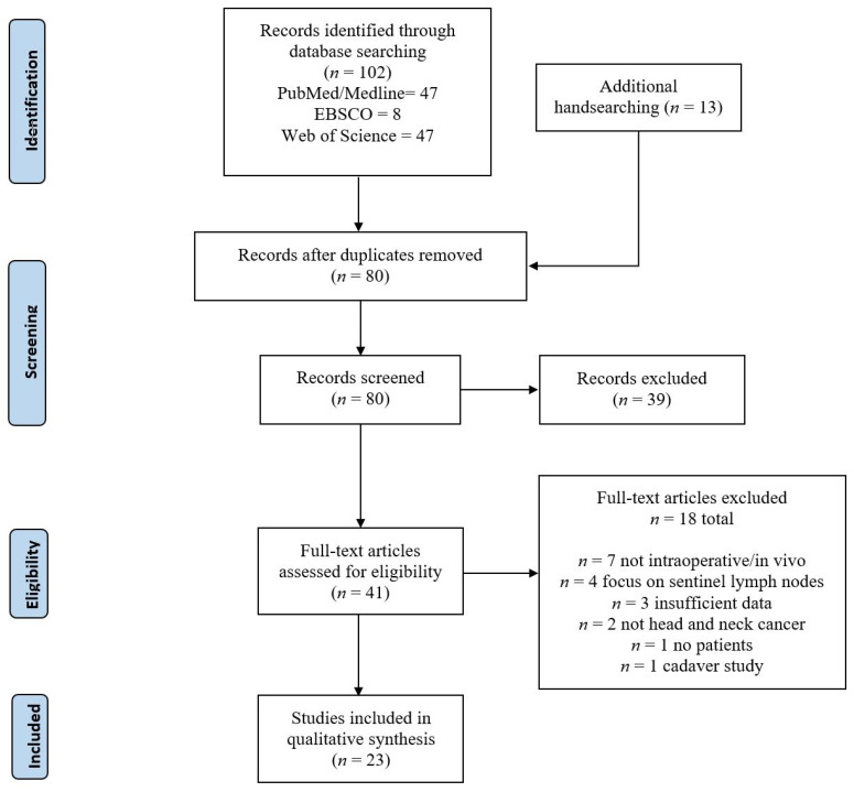
Surgical margin status is one of the strongest prognosticators in predicting patient outcomes in head and neck cancer, yet head and neck surgeons continue to face challenges in the accurate detection of these margins with the current standard of care. Novel intraoperative imaging modalities have demonstrated great promise for potentially increasing the accuracy and efficiency in surgical margin delineation. In this current study, we collated and analyzed various intraoperative imaging modalities utilized in head and neck cancer to evaluate their use in discriminating malignant from healthy tissues. The authors conducted a systematic database search through PubMed/Medline, Web of Science, and EBSCOhost (CINAHL). Study screening and data extraction were performed and verified by the authors, and more studies were added through handsearching. Here, intraoperative imaging modalities are described, including optical coherence tomography, narrow band imaging, autofluorescence, and fluorescent-tagged probe techniques. Available sensitivities and specificities in delineating cancerous from healthy tissues ranged from 83.0% to 100.0% and 79.2% to 100.0%, respectively, across the different imaging modalities. Many of these initial studies are in small sample sizes, with methodological differences that preclude more extensive quantitative comparison. Thus, there is impetus for future larger studies examining and comparing the efficacy of these intraoperative imaging technologies.
手术切缘状态是预测头颈癌患者预后的最强预后因素之一,但头颈外科医生在采用当前的治疗标准准确检测这些切缘时仍面临挑战。新型术中成像模式已显示出巨大的前景,有望提高手术切缘划定的准确性和效率。在本研究中,我们整理并分析了头颈癌中使用的各种术中成像模式,以评估它们在区分恶性组织和健康组织方面的应用。作者通过PubMed/Medline、科学网和EBSCOhost(护理学与健康领域数据库)进行了系统的数据库检索。研究筛选和数据提取由作者进行并验证,通过手工检索增加了更多研究。在此,描述了术中成像模式,包括光学相干断层扫描、窄带成像、自体荧光和荧光标记探针技术。在不同的成像模式中,区分癌组织和健康组织的可用灵敏度和特异性分别为83.0%至100.0%和79.2%至100.0%。许多这些初步研究样本量较小,存在方法学差异,无法进行更广泛的定量比较。因此,未来有必要开展更大规模的研究来检验和比较这些术中成像技术的疗效。