Baram Tamir, Oren Nino, Erlichman Nofar, Meshel Tsipi, Ben-Baruch Adit
The Shmunis School of Biomedicine and Cancer Research, George S. Wise Faculty of Life Sciences, Tel Aviv University, Tel Aviv 6997801, Israel.
Cancers (Basel). 2022 Jul 19;14(14):3513. doi: 10.3390/cancers14143513.
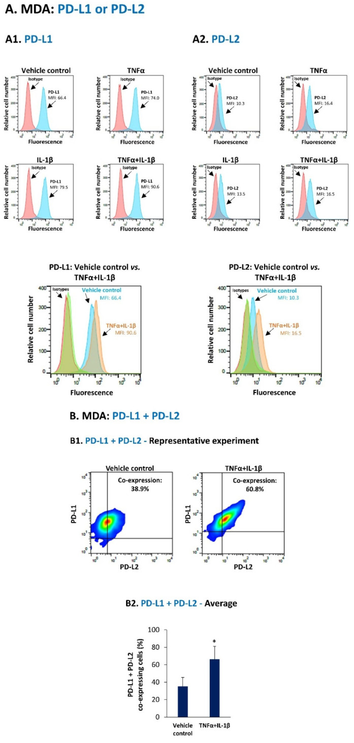
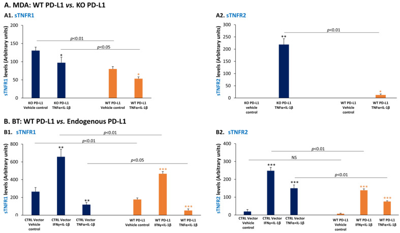
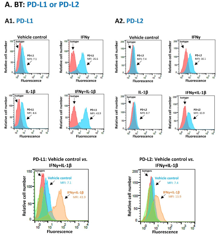
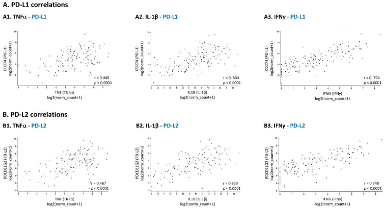
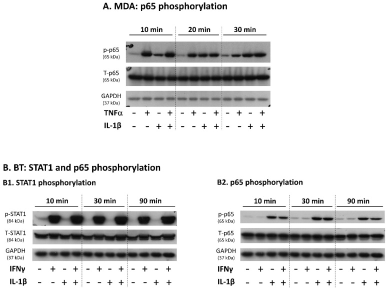
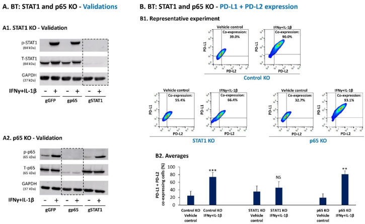
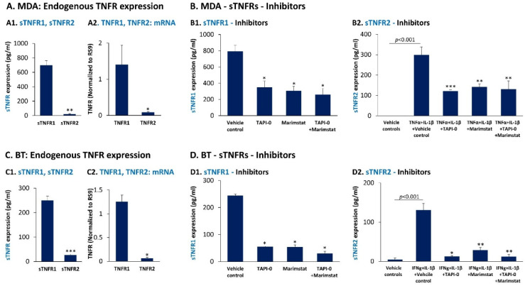
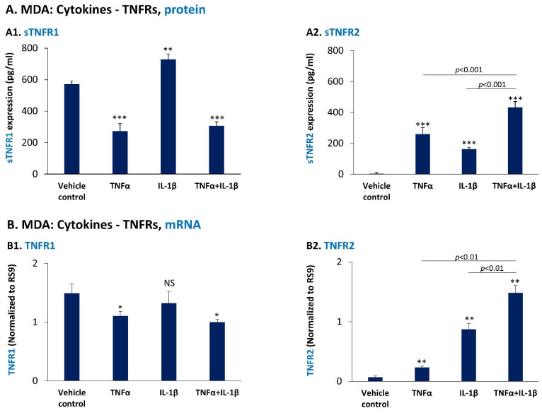
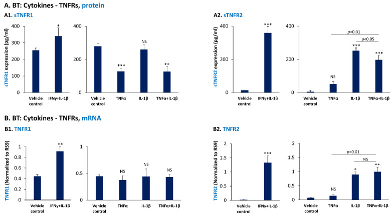
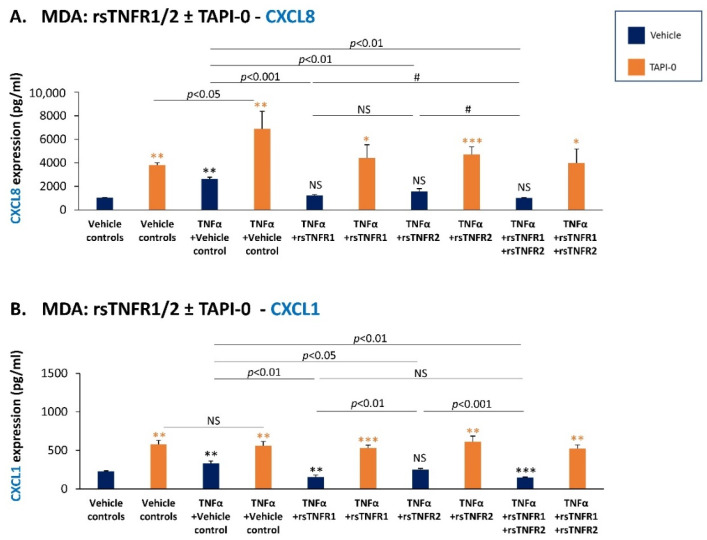
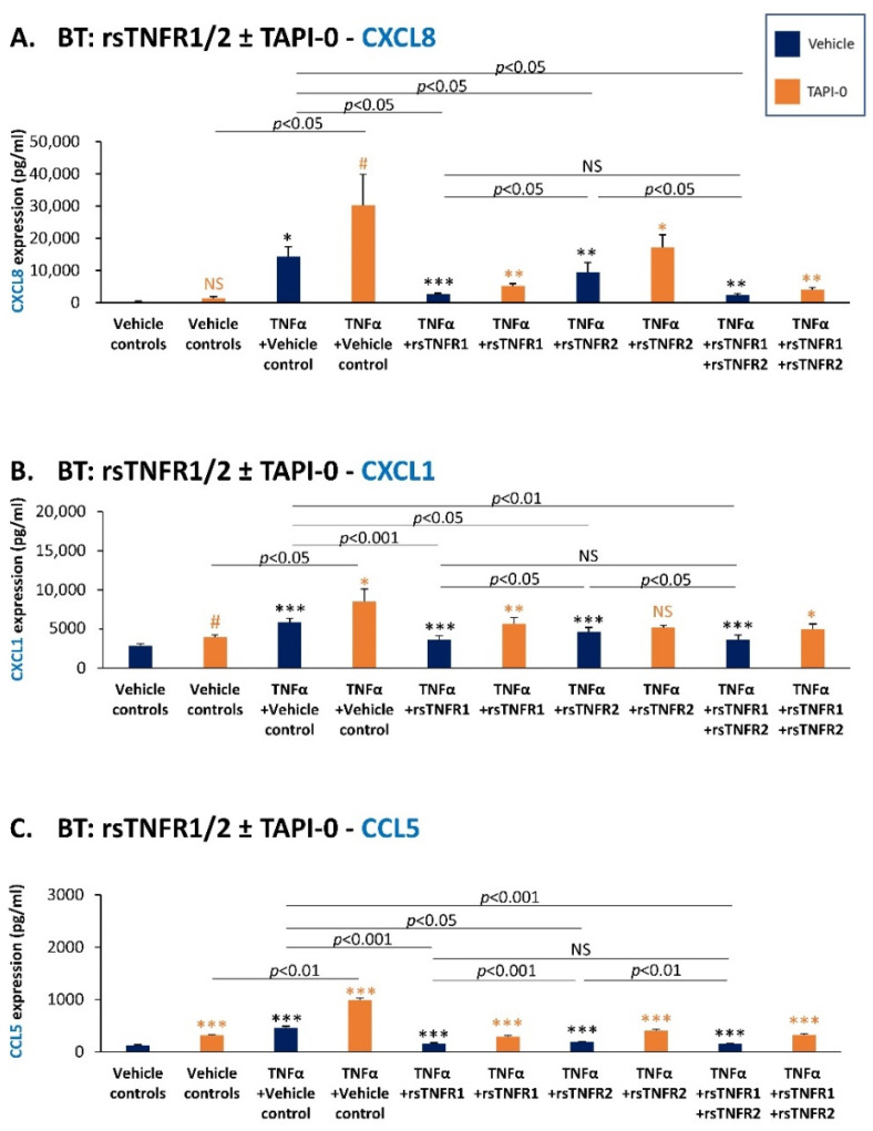
Pro-inflammatory cytokines play key roles in elevating cancer progression in triple-negative breast cancer (TNBC). We demonstrate that specific combinations between TNFα, IL-1β and IFNγ up-regulated the proportion of human TNBC cells co-expressing the inhibitory immune checkpoints PD-L1 and PD-L2: TNFα + IL-1β in MDA-MB-231 cells and IFNγ + IL-1β in BT-549 cells; in the latter cells, the process depended entirely on STAT1 activation, with no involvement of p65 (CRISPR-Cas9 experiments). Highly significant associations between the pro-inflammatory cytokines and PD-L1/PD-L2 expression were revealed in the TCGA dataset of basal-like breast cancer patients. In parallel, we found that the pro-inflammatory cytokines regulated the expression of the soluble receptors of tumor necrosis factor α (TNFα), namely sTNFR1 and sTNFR2; moreover, we revealed that sTNFR1 and sTNFR2 serve as anti-metastatic and protective factors in TNBC, reducing the TNFα-induced production of inflammatory pro-metastatic chemokines (CXCL8, CXCL1, CCL5) by TNBC cells. Importantly, we found that in the context of inflammatory stimulation and also without exposure to pro-inflammatory cytokines, elevated levels of PD-L1 have down-regulated the production of anti-tumor sTNFR1 and sTNFR2. These findings suggest that in addition to its immune-suppressive activities, PD-L1 may promote disease course in TNBC by inhibiting the protective effects of sTNFR1 and sTNFR2.
促炎细胞因子在三阴性乳腺癌(TNBC)的癌症进展中起关键作用。我们证明,TNFα、IL-1β和IFNγ之间的特定组合上调了共表达抑制性免疫检查点PD-L1和PD-L2的人TNBC细胞比例:MDA-MB-231细胞中的TNFα + IL-1β以及BT-549细胞中的IFNγ + IL-1β;在后者细胞中,该过程完全依赖于STAT1激活,而p65不参与(CRISPR-Cas9实验)。在基底样乳腺癌患者的TCGA数据集中,揭示了促炎细胞因子与PD-L1/PD-L2表达之间高度显著的关联。同时,我们发现促炎细胞因子调节肿瘤坏死因子α(TNFα)的可溶性受体sTNFR1和sTNFR2的表达;此外,我们揭示sTNFR1和sTNFR2在TNBC中作为抗转移和保护因子,减少TNBC细胞中TNFα诱导的促炎促转移趋化因子(CXCL8、CXCL1、CCL5)的产生。重要的是,我们发现,在炎症刺激的背景下以及未暴露于促炎细胞因子的情况下,PD-L1水平升高下调了抗肿瘤sTNFR1和sTNFR2的产生。这些发现表明,除了其免疫抑制活性外,PD-L1可能通过抑制sTNFR1和sTNFR2的保护作用来促进TNBC的病程发展。