Dinkel Johannes G, Lahmer Godehard, Mennecke Angelika, Hock Stefan W, Richter-Schmidinger Tanja, Fietkau Rainer, Distel Luitpold, Putz Florian, Dörfler Arnd, Schmidt Manuel A
Neuroradiologisches Institut des Universitätsklinikums Erlangen, Friedrich-Alexander-Universität Erlangen-Nürnberg (FAU), 91054 Erlangen, Germany.
Strahlenklinik des Universitätsklinikums Erlangen, Friedrich-Alexander-Universität Erlangen-Nürnberg (FAU), 91054 Erlangen, Germany.
Brain Sci. 2022 Jul 4;12(7):879. doi: 10.3390/brainsci12070879.
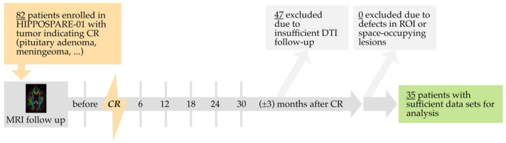
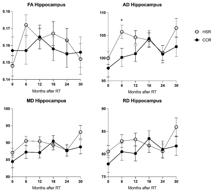
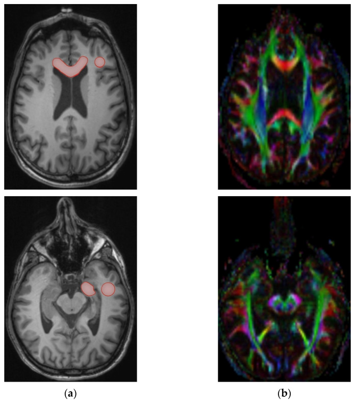
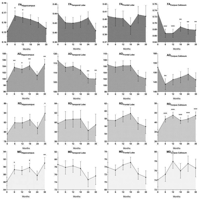
Hippocampal-sparing radiotherapy (HSR) is a promising approach to alleviate cognitive side effects following cranial radiotherapy. Microstructural brain changes after irradiation have been demonstrated using Diffusion Tensor Imaging (DTI). However, evidence is conflicting for certain parameters and anatomic structures. This study examines the effects of radiation on white matter and hippocampal microstructure using DTI and evaluates whether these may be mitigated using HSR. A total of 35 tumor patients undergoing a prospective randomized controlled trial receiving either conventional or HSR underwent DTI before as well as 6, 12, 18, 24, and 30 (±3) months after radiotherapy. Fractional Anisotropy (FA), Mean Diffusivity (MD), Axial Diffusivity (AD), and Radial Diffusivity (RD) were measured in the hippocampus (CA), temporal, and frontal lobe white matter (TL, FL), and corpus callosum (CC). Longitudinal analysis was performed using linear mixed models. Analysis of the entire patient collective demonstrated an overall FACC decrease and RDCC increase compared to baseline in all follow-ups; ADCC decreased after 6 months, and MDCC increased after 12 months (p ≤ 0.001, 0.001, 0.007, 0.018). ADTL decreased after 24 and 30 months (p ≤ 0.004, 0.009). Hippocampal FA increased after 6 and 12 months, driven by a distinct increase in ADCA and MDCA, with RDCA not increasing until 30 months after radiotherapy (p ≤ 0.011, 0.039, 0.005, 0.040, 0.019). Mean radiation dose correlated positively with hippocampal FA (p < 0.001). These findings may indicate complex pathophysiological changes in cerebral microstructures after radiation, insufficiently explained by conventional DTI models. Hippocampal microstructure differed between patients undergoing HSR and conventional cranial radiotherapy after 6 months with a higher ADCA in the HSR subgroup (p ≤ 0.034).
保留海马体的放射疗法(HSR)是一种有望减轻颅脑放疗后认知副作用的方法。使用扩散张量成像(DTI)已证实了放疗后大脑微观结构的变化。然而,某些参数和解剖结构的证据存在冲突。本研究使用DTI检查辐射对白质和海马体微观结构的影响,并评估HSR是否可以减轻这些影响。共有35名接受前瞻性随机对照试验的肿瘤患者,他们分别接受传统放疗或HSR,在放疗前以及放疗后6、12、18、24和30(±3)个月进行了DTI检查。在海马体(CA)、颞叶和额叶白质(TL、FL)以及胼胝体(CC)中测量了分数各向异性(FA)、平均扩散率(MD)、轴向扩散率(AD)和径向扩散率(RD)。使用线性混合模型进行纵向分析。对整个患者群体的分析表明,与基线相比,在所有随访中,CC的FACC总体下降,RDCC增加;6个月后ADCC下降,12个月后MDCC增加(p≤0.001、0.001、0.007、0.018)。24和30个月后ADTL下降(p≤0.004、0.009)。放疗后6和12个月海马体FA增加,这是由ADCA和MDCA的明显增加驱动的,直到放疗后30个月RDCA才增加(p≤0.011、0.039、0.005、0.040、0.019)。平均辐射剂量与海马体FA呈正相关(p<0.001)。这些发现可能表明放疗后大脑微观结构存在复杂的病理生理变化,传统DTI模型对此解释不足。6个月后,接受HSR和传统颅脑放疗的患者海马体微观结构不同,HSR亚组的ADCA更高(p≤0.034)。