• 文献检索
  • 文档翻译
  • 深度研究
  • 学术资讯
  • Suppr Zotero 插件Zotero 插件
  • 邀请有礼
  • 套餐&价格
  • 历史记录
应用&插件
Suppr Zotero 插件Zotero 插件浏览器插件Mac 客户端Windows 客户端微信小程序
定价
高级版会员购买积分包购买API积分包
服务
文献检索文档翻译深度研究API 文档MCP 服务
关于我们
关于 Suppr公司介绍联系我们用户协议隐私条款
关注我们

Suppr 超能文献

核心技术专利:CN118964589B侵权必究
粤ICP备2023148730 号-1Suppr @ 2026

文献检索

告别复杂PubMed语法,用中文像聊天一样搜索,搜遍4000万医学文献。AI智能推荐,让科研检索更轻松。

立即免费搜索

文件翻译

保留排版,准确专业,支持PDF/Word/PPT等文件格式,支持 12+语言互译。

免费翻译文档

深度研究

AI帮你快速写综述,25分钟生成高质量综述,智能提取关键信息,辅助科研写作。

立即免费体验

结节状和微结节状基底细胞癌亚型基于其形态结构及其与周围基质的相互作用而属于不同的肿瘤。

Nodular and Micronodular Basal Cell Carcinoma Subtypes Are Different Tumors Based on Their Morphological Architecture and Their Interaction with the Surrounding Stroma.

作者信息

Șerbănescu Mircea-Sebastian, Bungărdean Raluca Maria, Georgiu Carmen, Crișan Maria

机构信息

Department of Medical Informatics and Biostatistics, University of Medicine and Pharmacy of Craiova, 200349 Craiova, Romania.

Department of Pathology, Iuliu Haţieganu University of Medicine and Pharmacy, 400012 Cluj-Napoca, Romania.

出版信息

Diagnostics (Basel). 2022 Jul 5;12(7):1636. doi: 10.3390/diagnostics12071636.

DOI:10.3390/diagnostics12071636
PMID:35885545
原文链接:https://pmc.ncbi.nlm.nih.gov/articles/PMC9323345/
Abstract

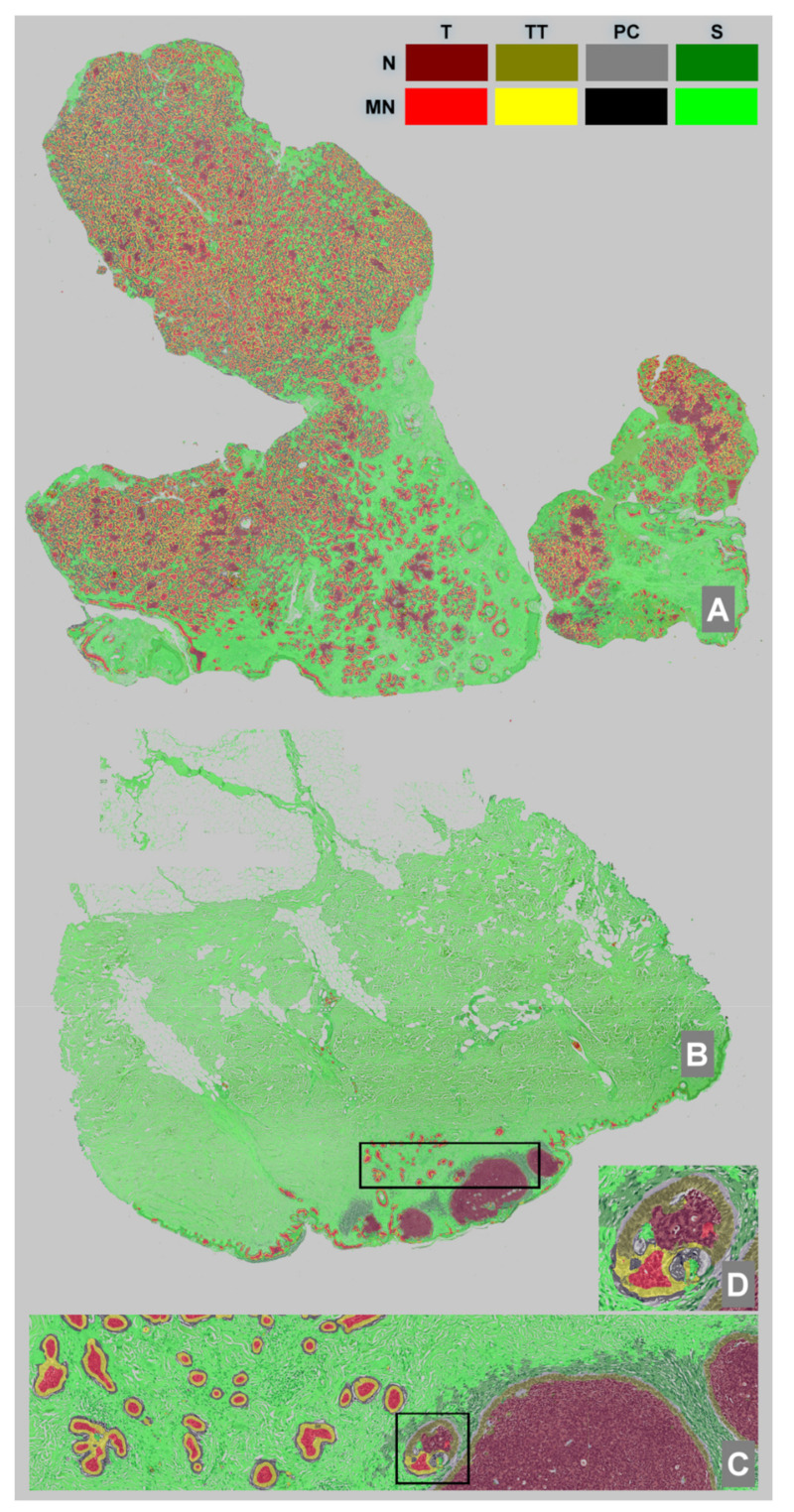
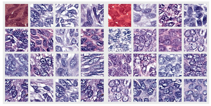
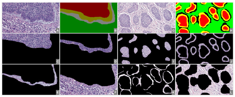
Basal cell carcinoma (BCC) is the most frequent cancer of the skin and comprises low-risk and high-risk subtypes. We selected a low-risk subtype, namely, nodular (N), and a high-risk subtype, namely, micronodular (MN), with the aim to identify differences between them using a classical morphometric approach through a gray-level co-occurrence matrix and histogram analysis, as well as an approach based on deep learning semantic segmentation. From whole-slide images, pathologists selected 216 N and 201 MN BCC images. The two groups were then manually segmented and compared based on four morphological areas: center of the BCC islands (tumor, T), peripheral palisading of the BCC islands (touching tumor, TT), peritumoral cleft (PC) and surrounding stroma (S). We found that the TT pattern varied the least, while the PC pattern varied the most between the two subtypes. The combination of two distinct analysis approaches yielded fresh insights into the characterization of BCC, and thus, we were able to describe two different morphological patterns for the T component of the two subtypes.

摘要

基底细胞癌(BCC)是最常见的皮肤癌,包括低风险和高风险亚型。我们选择了一种低风险亚型,即结节型(N),和一种高风险亚型,即微结节型(MN),旨在通过灰度共生矩阵和直方图分析的经典形态测量方法以及基于深度学习语义分割的方法来识别它们之间的差异。病理学家从全切片图像中选取了216张N型和201张MN型BCC图像。然后对这两组图像进行手动分割,并基于四个形态学区域进行比较:BCC岛中心(肿瘤,T)、BCC岛周边栅栏状结构(接触肿瘤,TT)、肿瘤周围裂隙(PC)和周围基质(S)。我们发现,在两种亚型之间,TT模式变化最小,而PC模式变化最大。两种不同分析方法的结合为BCC的特征描述提供了新的见解,因此,我们能够描述两种亚型T成分的两种不同形态模式。

https://cdn.ncbi.nlm.nih.gov/pmc/blobs/813f/9323345/5703d2fe8d8d/diagnostics-12-01636-g003.jpg
https://cdn.ncbi.nlm.nih.gov/pmc/blobs/813f/9323345/838aa5422af4/diagnostics-12-01636-g002.jpg
https://cdn.ncbi.nlm.nih.gov/pmc/blobs/813f/9323345/ff7dddab7686/diagnostics-12-01636-g001.jpg
https://cdn.ncbi.nlm.nih.gov/pmc/blobs/813f/9323345/5703d2fe8d8d/diagnostics-12-01636-g003.jpg
https://cdn.ncbi.nlm.nih.gov/pmc/blobs/813f/9323345/838aa5422af4/diagnostics-12-01636-g002.jpg
https://cdn.ncbi.nlm.nih.gov/pmc/blobs/813f/9323345/ff7dddab7686/diagnostics-12-01636-g001.jpg
https://cdn.ncbi.nlm.nih.gov/pmc/blobs/813f/9323345/5703d2fe8d8d/diagnostics-12-01636-g003.jpg

相似文献

1
Nodular and Micronodular Basal Cell Carcinoma Subtypes Are Different Tumors Based on Their Morphological Architecture and Their Interaction with the Surrounding Stroma.结节状和微结节状基底细胞癌亚型基于其形态结构及其与周围基质的相互作用而属于不同的肿瘤。
Diagnostics (Basel). 2022 Jul 5;12(7):1636. doi: 10.3390/diagnostics12071636.
2
Basal cell carcinoma progression correlates with host immune response and stromal alterations: a histologic analysis.基底细胞癌进展与宿主免疫反应及间质改变相关:一项组织学分析。
Am J Dermatopathol. 2006 Aug;28(4):293-307. doi: 10.1097/00000372-200608000-00002.
3
Micronodular basal cell carcinoma: a distinct subtype? Relationship with nodular and infiltrative basal cell carcinomas.微结节性基底细胞癌:一种独特的亚型?与结节性和浸润性基底细胞癌的关系。
J Dermatol. 2010 Jul;37(7):611-6. doi: 10.1111/j.1346-8138.2009.00772.x.
4
Differentiation and risk stratification of basal cell carcinoma with deep learning on histopathologic images and measuring nuclei and tumor microenvironment features.基于组织病理图像和测量细胞核及肿瘤微环境特征的深度学习方法对基底细胞癌的分化和危险分层。
Skin Res Technol. 2024 Jan;30(1):e13571. doi: 10.1111/srt.13571.
5
Basal cell carcinoma of the skin with mixed histomorphology: a comparative study.具有混合组织形态学的皮肤基底细胞癌:一项比较研究
Cesk Patol. 2016 Fall;52(4):222-226.
6
Increased immunoreactivity of membrane type-1 matrix metalloproteinase (MT1-MMP) and β-catenin in high-risk basal cell carcinoma.高风险基底细胞癌中膜型基质金属蛋白酶-1(MT1-MMP)和β-连环蛋白的免疫反应性增强。
Br J Dermatol. 2011 Dec;165(6):1197-204. doi: 10.1111/j.1365-2133.2011.10506.x.
7
Recognizing basal cell carcinoma on smartphone-captured digital histopathology images with a deep neural network.利用深度神经网络在智能手机拍摄的数字组织病理学图像上识别基底细胞癌。
Br J Dermatol. 2020 Mar;182(3):754-762. doi: 10.1111/bjd.18026. Epub 2019 Aug 22.
8
Micronodular basal cell carcinoma. A deceptive histologic subtype with frequent clinically undetected tumor extension.微结节型基底细胞癌。一种具有欺骗性的组织学亚型,临床上常有未被检测到的肿瘤扩展。
Arch Dermatol. 1996 Mar;132(3):295-8. doi: 10.1001/archderm.132.3.295.
9
In vivo diagnosis of basal cell carcinoma subtype by reflectance confocal microscopy.利用反射式共聚焦显微镜对基底细胞癌亚型进行体内诊断。
Dermatology. 2013;227(3):255-62. doi: 10.1159/000354762. Epub 2013 Oct 18.
10
Evaluation of Patched-1 Protein Expression Level in Low Risk and High Risk Basal Cell Carcinoma Subtypes.低风险和高风险基底细胞癌亚型中Patched-1蛋白表达水平的评估
Asian Pac J Cancer Prev. 2019 Sep 1;20(9):2851-2857. doi: 10.31557/APJCP.2019.20.9.2851.

引用本文的文献

1
Basal Cell Carcinoma: An Old Friend with Multiple Faces.基底细胞癌:一位有着多副面孔的老朋友。
Cancers (Basel). 2025 Mar 15;17(6):993. doi: 10.3390/cancers17060993.
2
Transfer Learning-Based Integration of Dual Imaging Modalities for Enhanced Classification Accuracy in Confocal Laser Endomicroscopy of Lung Cancer.基于迁移学习的双成像模态整合用于提高肺癌共聚焦激光内镜检查的分类准确性
Cancers (Basel). 2025 Feb 11;17(4):611. doi: 10.3390/cancers17040611.
3
Automatic Identification of Fetal Abdominal Planes from Ultrasound Images Based on Deep Learning.

本文引用的文献

1
Deep learning with transfer learning in pathology. Case study: classification of basal cell carcinoma.深度学习与迁移学习在病理学中的应用。案例研究:基底细胞癌分类。
Rom J Morphol Embryol. 2021 Oct-Dec;62(4):1017-1028. doi: 10.47162/RJME.62.4.14.
2
Laser Capture Microdissection in the Spatial Analysis of Epigenetic Modifications in Skin: A Comprehensive Review.激光捕获微切割在皮肤表观遗传修饰的空间分析中的应用:全面综述。
Oxid Med Cell Longev. 2022 Feb 9;2022:4127238. doi: 10.1155/2022/4127238. eCollection 2022.
3
Multiplexed nanomaterial-assisted laser desorption/ionization for pan-cancer diagnosis and classification.
基于深度学习的超声图像中胎儿腹部平面的自动识别
J Imaging Inform Med. 2025 Feb 5. doi: 10.1007/s10278-025-01409-6.
4
Editorial on Special Issue "Artificial Intelligence in Pathological Image Analysis".关于“病理图像分析中的人工智能”特刊的社论
Diagnostics (Basel). 2023 Feb 21;13(5):828. doi: 10.3390/diagnostics13050828.
多重纳米材料辅助激光解吸/电离用于泛癌诊断和分类。
Nat Commun. 2022 Feb 1;13(1):617. doi: 10.1038/s41467-021-26642-9.
4
'Retraction artefacts' in basal cell carcinomas do not result from fixation but likely arise by degradation of extracellular matrix during tumour growth.基底细胞癌中的“退缩伪像”并非由固定引起,而是可能在肿瘤生长过程中因细胞外基质降解而产生。
J Eur Acad Dermatol Venereol. 2022 Mar;36(3):e244-e247. doi: 10.1111/jdv.17776. Epub 2021 Nov 12.
5
Whole-slide margin control through deep learning in Mohs micrographic surgery for basal cell carcinoma.基于深度学习的全切片边缘控制在基底细胞癌 Mohs 显微外科手术中的应用。
Exp Dermatol. 2021 May;30(5):733-738. doi: 10.1111/exd.14306. Epub 2021 Mar 3.
6
Image Segmentation Using Deep Learning: A Survey.基于深度学习的图像分割技术综述。
IEEE Trans Pattern Anal Mach Intell. 2022 Jul;44(7):3523-3542. doi: 10.1109/TPAMI.2021.3059968. Epub 2022 Jun 3.
7
Deep learning in cancer pathology: a new generation of clinical biomarkers.深度学习在癌症病理学中的应用:新一代临床生物标志物。
Br J Cancer. 2021 Feb;124(4):686-696. doi: 10.1038/s41416-020-01122-x. Epub 2020 Nov 18.
8
Artificial neural networks and pathologists recognize basal cell carcinomas based on different histological patterns.人工神经网络和病理学家基于不同的组织学模式识别基底细胞癌。
Mod Pathol. 2021 May;34(5):895-903. doi: 10.1038/s41379-020-00712-7. Epub 2020 Nov 13.
9
TOF-SIMS imaging reveals tumor heterogeneity and inflammatory response markers in the microenvironment of basal cell carcinoma.TOF-SIMS 成像揭示了基底细胞癌微环境中的肿瘤异质性和炎症反应标志物。
Biointerphases. 2020 Aug 28;15(4):041012. doi: 10.1116/6.0000340.
10
Microdissection-An Essential Prerequisite for Spatial Cancer Omics.显微解剖——空间癌症组学的必要前提。
Proteomics. 2020 Sep;20(17-18):e2000077. doi: 10.1002/pmic.202000077. Epub 2020 Jul 6.