Yang Mei, Xiong Jun, Zou Qiang, Wang Xi, Hu Ke, Zhao Qingyan
Department of Cardiology, Renmin Hospital of Wuhan University, Wuhan, China.
Cardiovascular Research Institute, Wuhan University, Wuhan, China.
Front Cardiovasc Med. 2022 Jul 11;9:915903. doi: 10.3389/fcvm.2022.915903. eCollection 2022.
Macrophage polarization is an important regulatory mechanism of ventricular remodeling. Studies have shown that sinapic acid (SA) exerts an anti-inflammatory effect. However, the effect of SA on macrophages is still unclear.
The purpose of the study was to investigate the role of SA in macrophage polarization and ventricular remodeling after myocardial infarction (MI).
An MI model was established by ligating the left coronary artery. The rats with MI were treated with SA for 1 or 4 weeks after MI. The effect of SA on bone marrow-derived macrophages (BMDMs) was also observed .
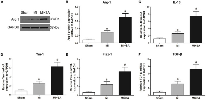
Cardiac systolic dysfunction was significantly improved after SA treatment. SA reduced MCP-1 and CCR2 expression and macrophage infiltration. SA decreased the levels of the inflammatory factors TNF-α, IL-1α, IL-1β, and iNOS and increased the levels of the M2 macrophage markers CD206, Arg-1, IL-10, Ym-1, Fizz-1, and TGF-β at 1 week after MI. SA significantly increased CD68/CD206 macrophage infiltration. Myocardial interstitial fibrosis and MMP-2 and MMP-9 levels were decreased, and the sympathetic nerve marker TH and nerve sprouting marker GAP43 were suppressed after SA treatment at 4 weeks after MI. The PPARγ level was notably upregulated after SA treatment. , SA also increased the expression of PPARγ mRNA in BMDMs and IL-4-treated BMDMs in a concentration-dependent manner. SA enhanced Arg1 and IL-10 expression in BMDMs, and the PPARγ antagonist GW9662 attenuated M2 macrophage marker expression.
Our results demonstrated that SA attenuated structural and neural remodeling by promoting macrophage M2 polarization PPARγ activation after MI.
巨噬细胞极化是心室重构的重要调节机制。研究表明,芥子酸(SA)具有抗炎作用。然而,SA对巨噬细胞的影响仍不清楚。
本研究旨在探讨SA在心肌梗死(MI)后巨噬细胞极化和心室重构中的作用。
通过结扎左冠状动脉建立MI模型。MI大鼠在MI后用SA治疗1或4周。还观察了SA对骨髓来源巨噬细胞(BMDM)的影响。
SA治疗后心脏收缩功能障碍明显改善。SA降低了MCP-1和CCR2的表达以及巨噬细胞浸润。SA在MI后1周降低了炎症因子TNF-α、IL-1α、IL-1β和iNOS的水平,并增加了M2巨噬细胞标志物CD206、Arg-1、IL-10、Ym-1、Fizz-1和TGF-β的水平。SA显著增加了CD68/CD206巨噬细胞浸润。MI后4周SA治疗后,心肌间质纤维化以及MMP-2和MMP-9水平降低,交感神经标志物TH和神经芽生标志物GAP43受到抑制。SA治疗后PPARγ水平显著上调。SA还以浓度依赖的方式增加了BMDM和IL-4处理的BMDM中PPARγ mRNA的表达。SA增强了BMDM中Arg1和IL-10的表达,PPARγ拮抗剂GW9662减弱了M2巨噬细胞标志物的表达。
我们的结果表明,SA通过促进MI后巨噬细胞M2极化和PPARγ激活减轻了结构和神经重构。