• 文献检索
  • 文档翻译
  • 深度研究
  • 学术资讯
  • Suppr Zotero 插件Zotero 插件
  • 邀请有礼
  • 套餐&价格
  • 历史记录
应用&插件
Suppr Zotero 插件Zotero 插件浏览器插件Mac 客户端Windows 客户端微信小程序
定价
高级版会员购买积分包购买API积分包
服务
文献检索文档翻译深度研究API 文档MCP 服务
关于我们
关于 Suppr公司介绍联系我们用户协议隐私条款
关注我们

Suppr 超能文献

核心技术专利:CN118964589B侵权必究
粤ICP备2023148730 号-1Suppr @ 2026

文献检索

告别复杂PubMed语法,用中文像聊天一样搜索,搜遍4000万医学文献。AI智能推荐,让科研检索更轻松。

立即免费搜索

文件翻译

保留排版,准确专业,支持PDF/Word/PPT等文件格式,支持 12+语言互译。

免费翻译文档

深度研究

AI帮你快速写综述,25分钟生成高质量综述,智能提取关键信息,辅助科研写作。

立即免费体验

钛酸钙或氧化铒纳米粒子对正常 HSF 细胞的细胞毒性和遗传毒性的评估。

Estimation of Calcium Titanate or Erbium Oxide Nanoparticles Induced Cytotoxicity and Genotoxicity in Normal HSF Cells.

机构信息

Zoology Department, Faculty of Science, Cairo University, Giza, Egypt.

Faculty of Biotechnology, October University for Modern Sciences and Arts, 6Th of October City, Egypt.

出版信息

Biol Trace Elem Res. 2023 May;201(5):2311-2318. doi: 10.1007/s12011-022-03354-9. Epub 2022 Jul 30.

DOI:10.1007/s12011-022-03354-9
PMID:35907160
原文链接:https://pmc.ncbi.nlm.nih.gov/articles/PMC10020245/
Abstract

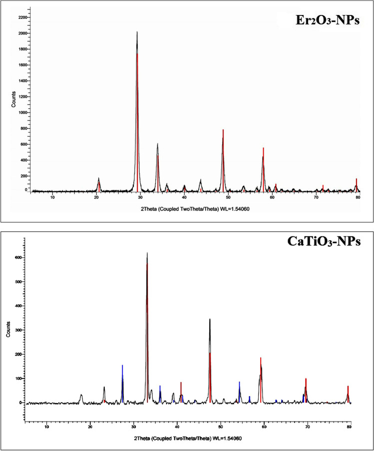
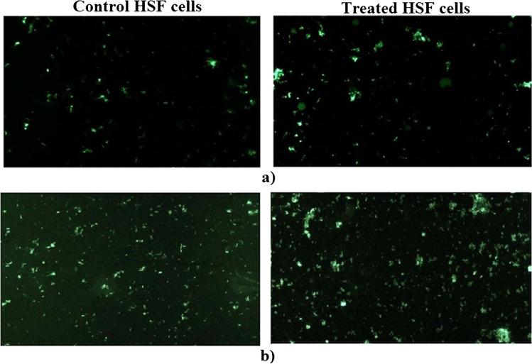
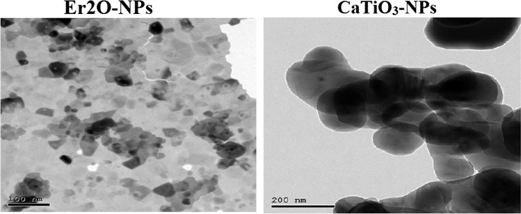
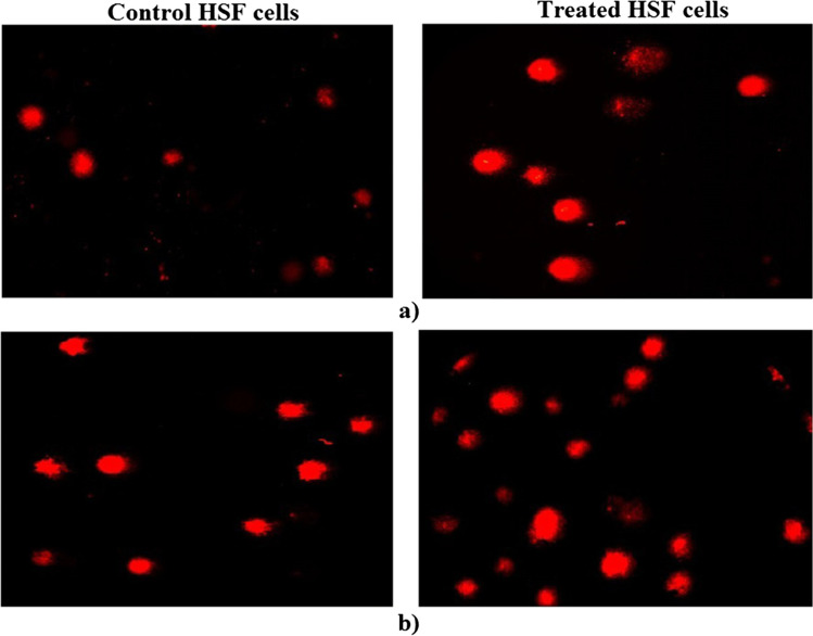
Extensive uses of calcium titanate nanoparticles (CaTiO-NPs) and erbium oxide nanoparticles (ErO-NPs) increase their release into the environment and human exposure, particularly through skin contact. However, there are almost no studies available on the effect of these nanoparticles on skin integrity. Therefore, this study was undertaken to estimate CaTiO-NP- or ErO-NP-induced cytotoxicity and genotoxicity in normal human skin fibroblast (HSF) cells. Cell viability was measured using sulforhodamine B (SRB) assay, while the level of DNA damage was detected using the alkaline comet assay. The intracellular levels of reactive oxygen species (ROS) as well as the expression level of p53, Bax, and Bcl2 genes were detected. Although the viability of HSF cells was non-markedly changed after 24 h, prolonged treatment with CaTiO3-NPs or Er2O3-NPs for 72 h induced concentration-dependent death of HSF cells. Treatment of normal HSF cells with IC50/72 h of CaTiO3-NPs or Er2O3-NPs did not cause marked changes in the intracellular level of ROS, DNA damage parameters, and expression levels of apoptosis genes compared to their values in the untreated HSF cells. We thus concluded that CaTiO3-NPs or Er2O3-NPs cause time- and concentration-dependent cytotoxicity toward normal HSF cells. However, safe and non-genotoxic effects were demonstrated by the apparent non-significant changes in intracellular ROS level, DNA integrity, and apoptotic genes' expression after exposure of normal HSF cells to nanoparticles. Thus, it is recommended that further studies be conducted to further understand the toxic and biological effects of CaTiO-NPs and Er2O3-NPs.

摘要

钛酸钙纳米粒子(CaTiO-NPs)和氧化铒纳米粒子(ErO-NPs)的广泛应用会导致它们释放到环境中并增加人体接触,尤其是通过皮肤接触。然而,目前几乎没有研究报道这些纳米粒子对皮肤完整性的影响。因此,本研究旨在评估正常人类皮肤成纤维细胞(HSF)中 CaTiO-NP 或 ErO-NP 诱导的细胞毒性和遗传毒性。使用磺酰罗丹明 B(SRB)测定法测量细胞活力,而使用碱性彗星试验检测 DNA 损伤水平。检测细胞内活性氧(ROS)水平以及 p53、Bax 和 Bcl2 基因的表达水平。虽然 HSF 细胞在 24 小时后活力没有明显变化,但长时间(72 小时)用 CaTiO3-NPs 或 Er2O3-NPs 处理会导致 HSF 细胞浓度依赖性死亡。与未经处理的 HSF 细胞相比,用 IC50/72 小时的 CaTiO3-NPs 或 Er2O3-NPs 处理正常 HSF 细胞不会导致细胞内 ROS 水平、DNA 损伤参数和凋亡基因表达水平发生明显变化。因此,我们得出结论,CaTiO3-NPs 或 Er2O3-NPs 会导致正常 HSF 细胞产生时间和浓度依赖性细胞毒性。然而,正常 HSF 细胞暴露于纳米粒子后,细胞内 ROS 水平、DNA 完整性和凋亡基因表达无明显变化,表明其具有安全的非遗传毒性作用。因此,建议进一步开展研究以进一步了解 CaTiO-NPs 和 Er2O3-NPs 的毒性和生物学效应。

https://cdn.ncbi.nlm.nih.gov/pmc/blobs/336e/10020245/bb1d8b0b07e4/12011_2022_3354_Fig5_HTML.jpg
https://cdn.ncbi.nlm.nih.gov/pmc/blobs/336e/10020245/add7bc55e881/12011_2022_3354_Fig1_HTML.jpg
https://cdn.ncbi.nlm.nih.gov/pmc/blobs/336e/10020245/b401804cd82f/12011_2022_3354_Fig2_HTML.jpg
https://cdn.ncbi.nlm.nih.gov/pmc/blobs/336e/10020245/854d1c3e3443/12011_2022_3354_Fig3_HTML.jpg
https://cdn.ncbi.nlm.nih.gov/pmc/blobs/336e/10020245/c6ecd1a87e29/12011_2022_3354_Fig4_HTML.jpg
https://cdn.ncbi.nlm.nih.gov/pmc/blobs/336e/10020245/bb1d8b0b07e4/12011_2022_3354_Fig5_HTML.jpg

相似文献

1
Estimation of Calcium Titanate or Erbium Oxide Nanoparticles Induced Cytotoxicity and Genotoxicity in Normal HSF Cells.钛酸钙或氧化铒纳米粒子对正常 HSF 细胞的细胞毒性和遗传毒性的评估。
Biol Trace Elem Res. 2023 May;201(5):2311-2318. doi: 10.1007/s12011-022-03354-9. Epub 2022 Jul 30.
2
Induction of oxidative DNA damage, cell cycle arrest and p53 mediated apoptosis by calcium titanate nanoparticles in MCF-7 breast cancer cells.钛酸钙纳米颗粒诱导MCF-7乳腺癌细胞发生氧化性DNA损伤、细胞周期阻滞及p53介导的细胞凋亡。
Cancer Cell Int. 2022 Nov 15;22(1):355. doi: 10.1186/s12935-022-02780-y.
3
Induction of ROS mediated genomic instability, apoptosis and G0/G1 cell cycle arrest by erbium oxide nanoparticles in human hepatic Hep-G2 cancer cells.氧化铒纳米颗粒通过诱导活性氧产生介导人类肝癌 Hep-G2 细胞基因组不稳定性、细胞凋亡和 G0/G1 期细胞周期阻滞。
Sci Rep. 2022 Sep 29;12(1):16333. doi: 10.1038/s41598-022-20830-3.
4
Zinc oxide nanoparticles exhibit cytotoxicity and genotoxicity through oxidative stress responses in human lung fibroblasts and .氧化锌纳米颗粒通过人肺成纤维细胞中的氧化应激反应表现出细胞毒性和基因毒性。
Int J Nanomedicine. 2017 Feb 28;12:1621-1637. doi: 10.2147/IJN.S124403. eCollection 2017.
5
Molybdenum nanoparticles-induced cytotoxicity, oxidative stress, G2/M arrest, and DNA damage in mouse skin fibroblast cells (L929).钼纳米颗粒诱导的小鼠皮肤成纤维细胞(L929)的细胞毒性、氧化应激、G2/M 期阻滞和 DNA 损伤。
Colloids Surf B Biointerfaces. 2015 Jan 1;125:73-81. doi: 10.1016/j.colsurfb.2014.11.014. Epub 2014 Nov 20.
6
Alleviation of calcium hydroxide nanoparticles induced genotoxicity and gastritis by coadministration of calcium titanate and yttrium oxide nanoparticles in mice.钙钛矿和氧化钇纳米粒子共给药缓解氢氧化钙纳米颗粒诱导的小鼠遗传毒性和胃炎。
Sci Rep. 2023 Dec 12;13(1):22011. doi: 10.1038/s41598-023-49303-x.
7
Cobalt oxide (Co3O4) nanoparticles induced genotoxicity in Chinese hamster lung fibroblast (V79) cells through modulation of reactive oxygen species.钴氧化物(Co3O4)纳米粒子通过调节活性氧引起中国仓鼠肺成纤维细胞(V79)的遗传毒性。
Mutagenesis. 2022 Apr 2;37(1):44-59. doi: 10.1093/mutage/geac005.
8
Yttrium Oxide nanoparticles induce cytotoxicity, genotoxicity, apoptosis, and ferroptosis in the human triple-negative breast cancer MDA-MB-231 cells.氧化钇纳米颗粒诱导人三阴性乳腺癌 MDA-MB-231 细胞的细胞毒性、遗传毒性、细胞凋亡和铁死亡。
BMC Cancer. 2023 Nov 27;23(1):1151. doi: 10.1186/s12885-023-11649-w.
9
Analysis of cytotoxicity and genotoxicity in a short-term dependent manner induced by a new titanium dioxide nanoparticle in murine fibroblast cells.分析新型二氧化钛纳米颗粒在小鼠成纤维细胞中短期依赖性诱导的细胞毒性和遗传毒性。
Toxicol Mech Methods. 2022 Mar;32(3):213-223. doi: 10.1080/15376516.2021.1994075. Epub 2021 Nov 9.
10
Genotoxicity and oxidative stress induction by calcium hydroxide, calcium titanate or/and yttrium oxide nanoparticles in mice.钙氢氧化物、钛酸钙和氧化钇纳米颗粒对小鼠的遗传毒性和氧化应激诱导作用。
Sci Rep. 2023 Nov 10;13(1):19633. doi: 10.1038/s41598-023-46522-0.

引用本文的文献

1
Induction of potent preferential cell death, severe DNA damage and p53-independent ROS-mediated mitochondrial apoptosis by CaTiONPs in HNO-97 tongue cancer cells.CaTiONPs在HNO-97舌癌细胞中诱导强效的选择性细胞死亡、严重的DNA损伤以及不依赖p53的活性氧介导的线粒体凋亡。
Naunyn Schmiedebergs Arch Pharmacol. 2025 Jun 4. doi: 10.1007/s00210-025-04323-4.
2
Erbium oxide nanoparticles induce potent cell death, genomic instability and ROS-mitochondrial dysfunction-mediated apoptosis in U937 lymphoma cells.氧化铒纳米颗粒可诱导U937淋巴瘤细胞发生强烈的细胞死亡、基因组不稳定以及活性氧-线粒体功能障碍介导的凋亡。
Naunyn Schmiedebergs Arch Pharmacol. 2025 Mar 12. doi: 10.1007/s00210-025-03962-x.
3

本文引用的文献

1
Enhanced Anticancer Performance of Eco-Friendly-Prepared Mo-ZnO/RGO Nanocomposites: Role of Oxidative Stress and Apoptosis.生态友好制备的Mo-ZnO/RGO纳米复合材料增强的抗癌性能:氧化应激和细胞凋亡的作用
ACS Omega. 2022 Feb 15;7(8):7103-7115. doi: 10.1021/acsomega.1c06789. eCollection 2022 Mar 1.
2
SnO-Doped ZnO/Reduced Graphene Oxide Nanocomposites: Synthesis, Characterization, and Improved Anticancer Activity via Oxidative Stress Pathway.SnO 掺杂 ZnO/还原氧化石墨烯纳米复合材料:通过氧化应激途径的合成、表征和提高抗癌活性。
Int J Nanomedicine. 2021 Jan 8;16:89-104. doi: 10.2147/IJN.S285392. eCollection 2021.
3
Induction of genotoxicity and differential alterations of p53 and inflammatory cytokines expression by acute oral exposure to bulk- or nano-calcium hydroxide particles in mice .
Potential Applications of Rare Earth Metal Nanoparticles in Biomedicine.
稀土金属纳米粒子在生物医学中的潜在应用
Pharmaceuticals (Basel). 2025 Jan 24;18(2):154. doi: 10.3390/ph18020154.
4
Calcium titanate nanoparticles-induced cytotoxicity, genotoxicity and oxidative stress in human non-small lung cancer cells.钛酸钙纳米颗粒对人非小细胞肺癌细胞的细胞毒性、遗传毒性和氧化应激作用
Sci Rep. 2025 Feb 21;15(1):6373. doi: 10.1038/s41598-025-89035-8.
5
YONPs induce selective cytotoxicity, genomic instability, oxidative stress and ROS mediated mitochondrial apoptosis in human epidermoid skin A-431 Cancer cells.YONPs在人表皮样皮肤A-431癌细胞中诱导选择性细胞毒性、基因组不稳定、氧化应激以及活性氧介导的线粒体凋亡。
Sci Rep. 2025 Jan 9;15(1):1543. doi: 10.1038/s41598-024-82376-w.
6
Exploring the cytotoxic and antioxidant properties of lanthanide-doped ZnO nanoparticles: a study with machine learning interpretation.探索镧系掺杂氧化锌纳米粒子的细胞毒性和抗氧化性能:基于机器学习解释的研究。
J Nanobiotechnology. 2024 Nov 10;22(1):687. doi: 10.1186/s12951-024-02957-9.
7
Yttrium oxide nanoparticles ameliorates calcium hydroxide and calcium titanate nanoparticles induced genomic DNA and mitochondrial damage, ROS generation and inflammation.氧化钇纳米颗粒可改善氢氧化钙和钛酸钙纳米颗粒引起的基因组 DNA 和线粒体损伤、ROS 生成和炎症。
Sci Rep. 2024 Jun 6;14(1):13015. doi: 10.1038/s41598-024-62877-4.
8
Alleviation of calcium hydroxide nanoparticles induced genotoxicity and gastritis by coadministration of calcium titanate and yttrium oxide nanoparticles in mice.钙钛矿和氧化钇纳米粒子共给药缓解氢氧化钙纳米颗粒诱导的小鼠遗传毒性和胃炎。
Sci Rep. 2023 Dec 12;13(1):22011. doi: 10.1038/s41598-023-49303-x.
9
Genotoxicity and oxidative stress induction by calcium hydroxide, calcium titanate or/and yttrium oxide nanoparticles in mice.钙氢氧化物、钛酸钙和氧化钇纳米颗粒对小鼠的遗传毒性和氧化应激诱导作用。
Sci Rep. 2023 Nov 10;13(1):19633. doi: 10.1038/s41598-023-46522-0.
10
Induction of oxidative DNA damage, cell cycle arrest and p53 mediated apoptosis by calcium titanate nanoparticles in MCF-7 breast cancer cells.钛酸钙纳米颗粒诱导MCF-7乳腺癌细胞发生氧化性DNA损伤、细胞周期阻滞及p53介导的细胞凋亡。
Cancer Cell Int. 2022 Nov 15;22(1):355. doi: 10.1186/s12935-022-02780-y.
急性经口摄入块状或纳米级氢氧化钙颗粒诱导小鼠遗传毒性及对 p53 和炎症细胞因子表达的差异改变。
Toxicol Mech Methods. 2021 Mar;31(3):169-181. doi: 10.1080/15376516.2020.1850961. Epub 2020 Dec 22.
4
DNA Damage/Repair Management in Cancers.癌症中的DNA损伤/修复管理
Cancers (Basel). 2020 Apr 23;12(4):1050. doi: 10.3390/cancers12041050.
5
Induction of fetal abnormalities and genotoxicity by molybdenum nanoparticles in pregnant female mice and fetuses.钼纳米颗粒在怀孕雌性小鼠及其胎儿中诱导的胎儿畸形和遗传毒性。
Environ Sci Pollut Res Int. 2020 Jul;27(19):23950-23962. doi: 10.1007/s11356-020-08137-0. Epub 2020 Apr 17.
6
Size, Surface Functionalization, and Genotoxicity of Gold Nanoparticles In Vitro.金纳米颗粒的尺寸、表面功能化及体外遗传毒性
Nanomaterials (Basel). 2020 Feb 6;10(2):271. doi: 10.3390/nano10020271.
7
Estimation of genomic instability and mitochondrial DNA damage induction by acute oral administration of calcium hydroxide normal- and nano- particles in mice.急性经口给予钙石灰纳米和常规粒子对小鼠基因组不稳定性和线粒体 DNA 损伤的评估。
Toxicol Lett. 2019 Apr;304:1-12. doi: 10.1016/j.toxlet.2018.12.012. Epub 2019 Jan 4.
8
Oxidative stress mediated cytotoxicity and apoptosis response of bismuth oxide (BiO) nanoparticles in human breast cancer (MCF-7) cells.氧化应激介导的氧化铋纳米粒子对人乳腺癌(MCF-7)细胞的细胞毒性和凋亡反应。
Chemosphere. 2019 Feb;216:823-831. doi: 10.1016/j.chemosphere.2018.10.214. Epub 2018 Oct 31.
9
Fingolimod interrupts the cross talk between estrogen metabolism and sphingolipid metabolism within prostate cancer cells.芬戈莫德中断了前列腺癌细胞内雌激素代谢和鞘脂代谢之间的串扰。
Toxicol Lett. 2018 Jul;291:77-85. doi: 10.1016/j.toxlet.2018.04.008. Epub 2018 Apr 11.
10
Comparison of cytotoxicity and genotoxicity effects of silver nanoparticles on human cervix and breast cancer cell lines.银纳米颗粒对人宫颈和乳腺癌细胞系的细胞毒性和遗传毒性作用比较。
Hum Exp Toxicol. 2017 Sep;36(9):931-948. doi: 10.1177/0960327116675206. Epub 2016 Nov 4.