Department of Radiology, Hallym University Sacred Heart Hospital, Anyang, Republic of Korea.
Public Health Wales Microbiology Cardiff, UHW, Cardiff CF14 4XW, UK.
Clin Imaging. 2022 Oct;90:11-18. doi: 10.1016/j.clinimag.2022.07.003. Epub 2022 Jul 23.
Common CT abnormalities of pulmonary aspergillosis represent a cavity with air-meniscus sign, nodule, mass, and consolidation having an angio-invasive pattern. This study aims to conduct a systematic review and an individual patient-level image analysis of CT findings of COVID-19-associated pulmonary aspergillosis (CAPA).
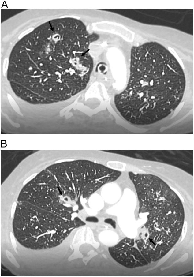
A systematic literature search was conducted to identify studies reporting CT findings of CAPA as of January 7, 2021. We summarized study-level clinical and CT findings of CAPA and collected individual patient CT images by inviting corresponding authors. The CT findings were categorized into four groups: group 1, typical appearance of COVID-19; group 2, indeterminate appearance of COVID-19; group 3, atypical for COVID-19 without cavities; and group 4, atypical for COVID-19 with cavities. In group 2, cases had only minor discrepant findings including solid nodules, isolated airspace consolidation with negligible ground-glass opacities, centrilobular micronodules, bronchial abnormalities, and cavities.
The literature search identified 89 patients from 25 studies, and we collected CT images from 35 CAPA patients (mean age 62.4 ± 14.6 years; 21 men): group 1, thirteen patients (37.1%); group 2, eight patients (22.9%); group 3, six patients (17.1%); and group 4, eight patients (22.9%). Eight of the 14 patients (57.1%) with an atypical appearance had bronchial abnormalities, whereas only one (7.1%) had an angio-invasive fungal pattern. In the study-level analysis, cavities were reported in 12 of 54 patients (22.2%).
CAPA can frequently manifest as COVID-19 pneumonia without common CT abnormalities of pulmonary aspergillosis. If abnormalities exist on CT images, CAPA may frequently accompany bronchial abnormalities.
肺部曲霉菌病的常见 CT 异常表现为具有气新月征的空洞、结节、肿块和实变,呈血管侵袭模式。本研究旨在对 COVID-19 相关肺曲霉菌病(CAPA)的 CT 表现进行系统回顾和个体患者水平的图像分析。
系统检索了截至 2021 年 1 月 7 日报告 CAPA CT 表现的研究。我们总结了 CAPA 的研究水平临床和 CT 表现,并通过邀请通讯作者收集了个体患者的 CT 图像。将 CT 表现分为四组:组 1,COVID-19 的典型表现;组 2,COVID-19 的不确定表现;组 3,无空洞的 COVID-19 不典型表现;组 4,有空洞的 COVID-19 不典型表现。在组 2 中,仅发现少数不一致的表现,包括实性结节、孤立性空气空间实变伴可忽略的磨玻璃影、小叶中心微结节、支气管异常和空洞。
文献检索从 25 项研究中确定了 89 名患者,我们从 35 名 CAPA 患者(平均年龄 62.4 ± 14.6 岁;21 名男性)中收集了 CT 图像:组 1,13 名患者(37.1%);组 2,8 名患者(22.9%);组 3,6 名患者(17.1%);组 4,8 名患者(22.9%)。14 名非典型表现患者中有 8 名(57.1%)存在支气管异常,而仅有 1 名(7.1%)存在血管侵袭性真菌模式。在研究水平分析中,54 名患者中有 12 名(22.2%)报告有空洞。
CAPA 常表现为 COVID-19 肺炎,无肺部曲霉菌病的常见 CT 异常。如果 CT 图像存在异常,CAPA 可能常伴有支气管异常。