Fischer Tim, El Baz Yassir, Graf Nicole, Wildermuth Simon, Leschka Sebastian, Kleger Gian-Reto, Pietsch Urs, Frischknecht Manuel, Scanferla Giulia, Strahm Carol, Wälti Stephan, Dietrich Tobias Johannes, Albrich Werner C
Division of Radiology and Nuclear Medicine, St. Gallen Cantonal Hospital, 9007 St. Gallen, Switzerland.
Clinical Trials Unit, St. Gallen Cantonal Hospital, 9007 St. Gallen, Switzerland.
Diagnostics (Basel). 2022 May 11;12(5):1201. doi: 10.3390/diagnostics12051201.
COVID-19 superinfection by Aspergillus (COVID-19-associated aspergillosis, CAPA) is increasingly observed due to increased awareness and use of corticosteroids. The aim of this study is to compare clinical and imaging features between COVID-19 patients with and without associated pulmonary aspergillosis.
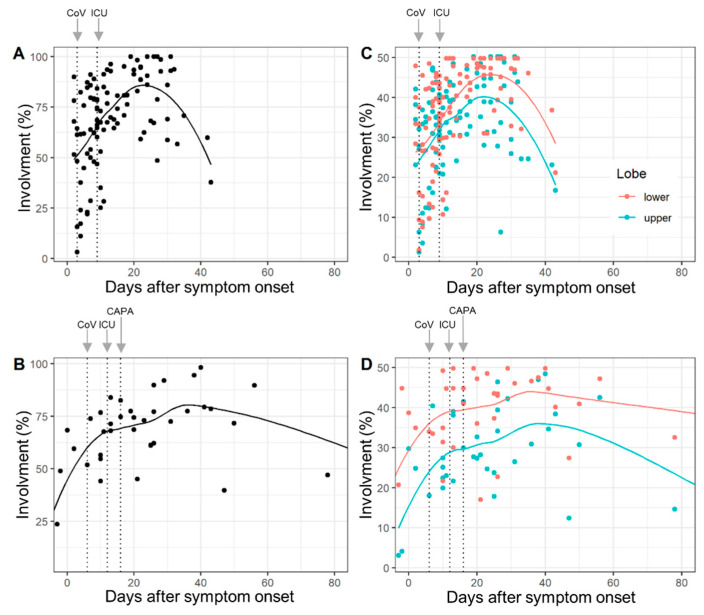
In this case-control study, hospitalized patients between March 2020 and March 2021 were evaluated. Two observers independently compared 105 chest CTs of 52 COVID-19 patients without pulmonary aspergillosis to 40 chest CTs of 13 CAPA patients. The following features were evaluated: lung involvement, predominant main pattern (ground glass opacity, crazy paving, consolidation) and additional lung and chest findings. Chronological changes in the abnormal extent upon CT and chronological changes in the main patterns were compared with mixed models. Patient-wise comparisons of additional features and demographic and clinical data were performed using Student's t-test, Chi-squared test, Fisher's exact tests and Wilcoxon rank-sum tests.
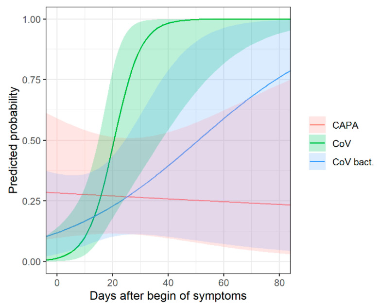
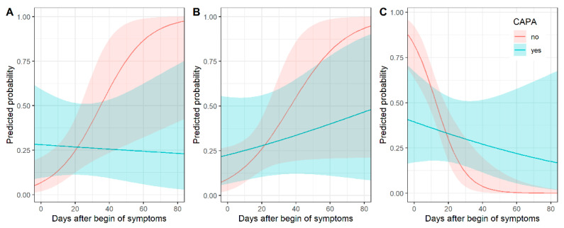
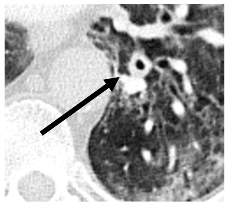
Compared to COVID-19 patients without pulmonary aspergillosis, CAPA patients were older (mean age (±SD): 70.3 (±7.8) versus 63.5 (±9.5) years ( = 0.01). The time-dependent evolution rates for consolidation ( = 0.02) and ground glass ( = 0.006) differed. In early COVID-19 disease, consolidation was associated with CAPA, whereas ground glass was less common. Chronological changes in the abnormal extent upon CT did not differ ( = 0.29). Regardless of the time point, bronchial wall thickening was observed more frequently in CAPA patients ( = 0.03).
CAPA patients showed a tendency for consolidation in early COVID-19 disease. Bronchial wall thickening and higher patient age were associated with CAPA. The overall lung involvement was similar between both groups.
由于对皮质类固醇的认识和使用增加,越来越多地观察到曲霉对新型冠状病毒肺炎(COVID-19)的重叠感染(与COVID-19相关的曲霉病,CAPA)。本研究的目的是比较合并和未合并肺部曲霉病的COVID-19患者的临床和影像学特征。
在这项病例对照研究中,对2020年3月至2021年3月期间住院的患者进行了评估。两名观察者独立地将52例无肺部曲霉病的COVID-19患者的105份胸部CT与13例CAPA患者的40份胸部CT进行比较。评估了以下特征:肺部受累情况、主要主要模式(磨玻璃影、铺路石样改变、实变)以及其他肺部和胸部表现。使用混合模型比较CT上异常范围的时间变化和主要模式的时间变化。使用学生t检验、卡方检验、费舍尔精确检验和威尔科克森秩和检验对其他特征以及人口统计学和临床数据进行患者层面的比较。
与无肺部曲霉病的COVID-19患者相比,CAPA患者年龄更大(平均年龄(±标准差):70.3(±7.8)岁对63.5(±9.5)岁(P = 0.01)。实变(P = 0.02)和磨玻璃影(P = 0.006)的时间依赖性演变率不同。在COVID-19疾病早期,实变与CAPA相关,而磨玻璃影较少见。CT上异常范围的时间变化无差异(P = 0.29)。无论时间点如何,CAPA患者中支气管壁增厚更为常见(P = 0.03)。
CAPA患者在COVID-19疾病早期有实变倾向。支气管壁增厚和患者年龄较大与CAPA相关。两组的总体肺部受累情况相似。