Department of Rheumatology, The First Affiliated Hospital, Sun Yat-sen University, Guangzhou, China.
Department of Pediatrics, The First Affiliated Hospital, Sun Yat-sen University, Guangzhou, China.
Clin Transl Med. 2022 Aug;12(8):e999. doi: 10.1002/ctm2.999.
T helper cells in patients with autoimmune disease of idiopathic inflammatory myopathies (IIM) are characterized with the proinflammatory phenotypes. The underlying mechanisms remain unknown.
RNA sequencing was performed for differential expression genes. Gene expression in CD4 T-cells was confirmed by quantitative real-time PCR. CD4 T-cells from IIM patients or healthy controls were evaluated for metabolic activities by Seahorse assay. Glucose uptake, T-cell proliferation and differentiation were evaluated and measured by flow cytometry. Human CD4 T-cells treated with iron chelators or Pfkfb4 siRNA were measured for glucose metabolism, proliferation and differentiation. Signalling pathway activation was evaluated by western blot and flow cytometry. Mouse model of experimental autoimmune myositis (EAM) were induced and treated with iron chelator or rapamycin. CD4 T-cell differentiation and muscle inflammation in the EAM mice were evaluated.
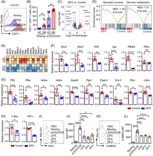
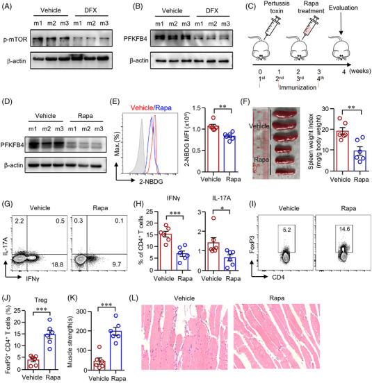
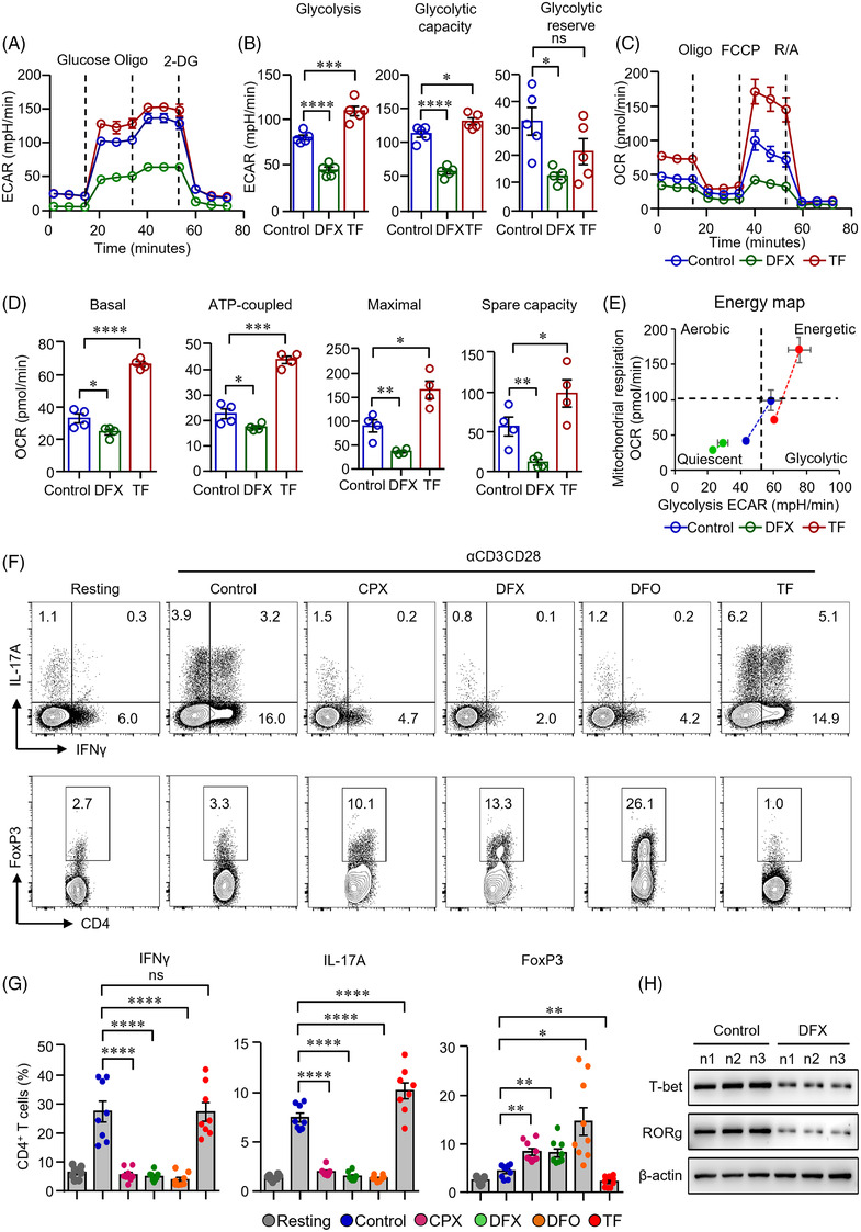
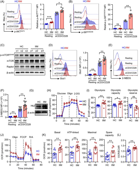
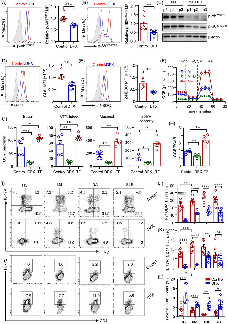
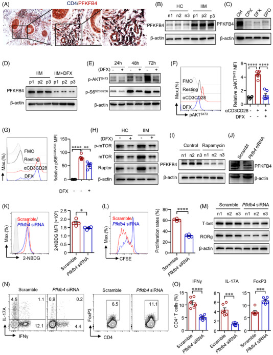
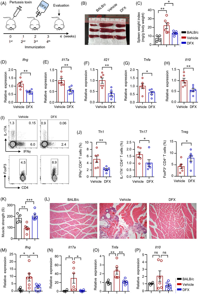
RNA-sequencing analysis revealed that iron was involved with glucose metabolism and CD4 T-cell differentiation. IIM patient-derived CD4 T-cells showed enhanced glycolysis and mitochondrial respiration, which was inhibited by iron chelation. CD4 T-cells from patients with IIM was proinflammatory and iron chelation suppressed the differentiation of interferon gamma (IFNγ)- and interleukin (IL)-17A-producing CD4 T-cells, which resulted in an increased percentage of regulatory T (Treg) cells. Mechanistically, iron promoted glucose metabolism by an upregulation of PFKFB4 through AKT-mTOR signalling pathway. Notably, the knockdown of Pfkfb4 decreased glucose influx and thus suppressed the differentiation of IFNγ- and IL-17A-producing CD4 T-cells. In vivo, iron chelation inhibited mTOR signalling pathway and reduced PFKFB4 expression in CD4 T-cells, resulting in reduced proinflammatory IFNγ- and IL-17A-producing CD4 T-cells and increased Foxp3 Treg cells, leading to ameliorated muscle inflammation.
Iron directs CD4 T-cells into a proinflammatory phenotype by enhancing glucose metabolism. Therapeutic targeting of iron metabolism should have the potential to normalize glucose metabolism in CD4 T-cells and reverse their proinflammatory phenotype in IIM.
特发性炎性肌病(IIM)自身免疫性疾病患者的辅助性 T 细胞表现出促炎表型。其潜在机制尚不清楚。
进行差异表达基因的 RNA 测序。通过实时定量 PCR 确认 CD4 T 细胞中的基因表达。通过 Seahorse 测定评估来自 IIM 患者或健康对照者的 CD4 T 细胞的代谢活性。通过流式细胞术评估和测量葡萄糖摄取、T 细胞增殖和分化。用铁螯合剂或 Pfkfb4 siRNA 处理人 CD4 T 细胞,以测量葡萄糖代谢、增殖和分化。通过 Western blot 和流式细胞术评估信号通路激活。用铁螯合剂或雷帕霉素诱导并处理实验性自身免疫性肌炎(EAM)小鼠模型。评估 EAM 小鼠中的 CD4 T 细胞分化和肌肉炎症。
RNA 测序分析表明,铁参与葡萄糖代谢和 CD4 T 细胞分化。IIM 患者来源的 CD4 T 细胞表现出增强的糖酵解和线粒体呼吸,这可被铁螯合所抑制。来自 IIM 患者的 CD4 T 细胞呈促炎性,铁螯合抑制干扰素 γ(IFNγ)和白细胞介素(IL)-17A 产生的 CD4 T 细胞的分化,导致调节性 T(Treg)细胞的百分比增加。在机制上,铁通过 AKT-mTOR 信号通路上调 PFKFB4 来促进葡萄糖代谢。值得注意的是,Pfkfb4 的敲低减少了葡萄糖流入,从而抑制了 IFNγ 和 IL-17A 产生的 CD4 T 细胞的分化。在体内,铁螯合抑制 mTOR 信号通路并降低 CD4 T 细胞中的 Pfkfb4 表达,导致减少促炎性 IFNγ 和 IL-17A 产生的 CD4 T 细胞和增加 Foxp3 Treg 细胞,从而改善肌肉炎症。
铁通过增强葡萄糖代谢将 CD4 T 细胞导向促炎表型。靶向铁代谢的治疗方法有可能使 CD4 T 细胞的葡萄糖代谢正常化并逆转其在 IIM 中的促炎表型。