Medical Imaging Research Center, Shiraz University of Medical Sciences, Shiraz, Iran.
Ongil, 79 D3, Sivaya Nagar, Reddiyur Alagapuram, Salem, India.
Iran J Med Sci. 2022 Jul;47(4):338-349. doi: 10.30476/IJMS.2021.90665.2165.
The present study aimed to evaluate the effectiveness of ultra-low-dose (ULD) chest computed tomography (CT) in comparison with the routine dose (RD) CT images in detecting lung lesions related to COVID-19.
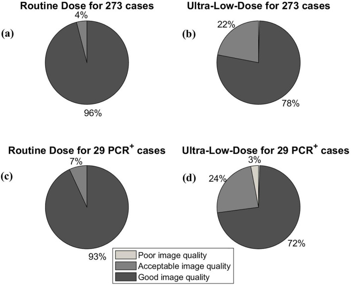
A prospective study was conducted during April-September 2020 at Shahid Faghihi Hospital affiliated with Shiraz University of Medical Sciences, Shiraz, Iran. In total, 273 volunteers with suspected COVID-19 participated in the study and successively underwent RD-CT and ULD-CT chest scans. Two expert radiologists qualitatively evaluated the images. Dose assessment was performed by determining volume CT dose index, dose length product, and size-specific dose estimate. Data analysis was performed using a ranking test and kappa coefficient (κ). P<0.05 was considered statistically significant.
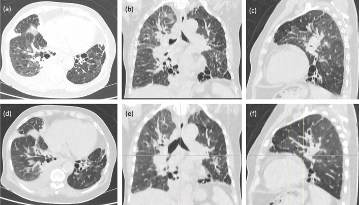
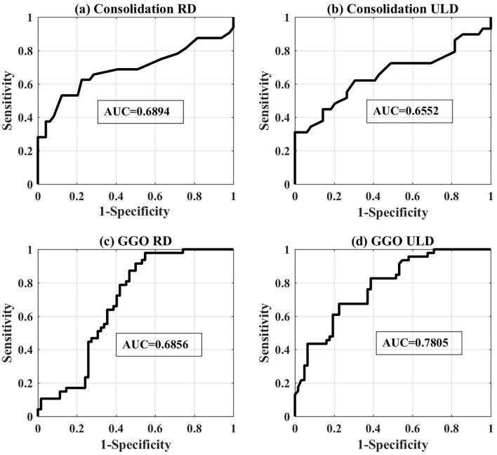
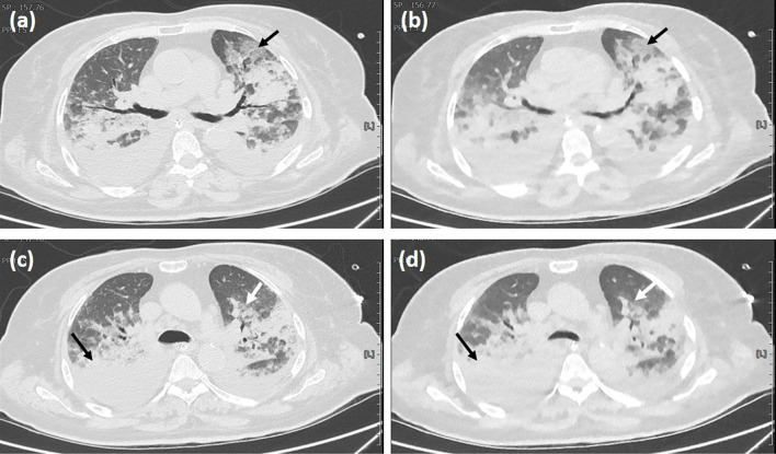
Lung lesions could be detected with both RD-CT and ULD-CT images in patients with suspected or confirmed COVID-19 (κ=1.0, P=0.016). The estimated effective dose for the RD-CT protocol was 22-fold higher than in the ULD-CT protocol. In the case of the ULD-CT protocol, sensitivity, specificity, accuracy, and positive predictive value for the detection of consolidation were 60%, 83%, 80%, and 20%, respectively. Comparably, in the case of RD-CT, these percentages for the detection of ground-glass opacity (GGO) were 62%, 66%, 66%, and 18%, respectively. Assuming the result of real-time polymerase chain reaction as true-positive, analysis of the receiver-operating characteristic curve for GGO detected using the ULD-CT protocol showed a maximum area under the curve of 0.78.
ULD-CT, with 94% dose reduction, can be an alternative to RD-CT to detect lung lesions for COVID-19 diagnosis and follow-up.An earlier preliminary report of a similar work with a lower sample size was submitted to the arXive as a preprint. The preprint is cited as: https://arxiv.org/abs/2005.03347.
本研究旨在评估超低剂量(ULD)胸部计算机断层扫描(CT)与常规剂量(RD)CT 图像在检测与 COVID-19 相关的肺部病变方面的效果。
本前瞻性研究于 2020 年 4 月至 9 月在伊朗设拉子谢里夫医科大学附属沙希德法希希医院进行。共有 273 名疑似 COVID-19 的志愿者参加了该研究,并相继接受了 RD-CT 和 ULD-CT 胸部扫描。两名专家放射科医生对图像进行了定性评估。通过确定容积 CT 剂量指数、剂量长度乘积和大小特异性剂量估计值来进行剂量评估。使用排序检验和 κ 系数(κ)进行数据分析。P<0.05 被认为具有统计学意义。
疑似或确诊 COVID-19 患者的 RD-CT 和 ULD-CT 图像均可检测到肺部病变(κ=1.0,P=0.016)。RD-CT 方案的估计有效剂量比 ULD-CT 方案高 22 倍。在 ULD-CT 方案中,检测实变的敏感性、特异性、准确性和阳性预测值分别为 60%、83%、80%和 20%。相比之下,在 RD-CT 方案中,检测磨玻璃密度(GGO)的这些百分比分别为 62%、66%、66%和 18%。假设实时聚合酶链反应的结果为真阳性,分析 ULD-CT 方案检测到的 GGO 的受试者工作特征曲线显示曲线下面积的最大值为 0.78。
ULD-CT 可作为 RD-CT 的替代方案,用于 COVID-19 诊断和随访时检测肺部病变,剂量降低 94%。一项类似工作的早期初步报告,样本量较小,已作为预印本提交给 arXive。该预印本被引述为:https://arxiv.org/abs/2005.03347。