He Michelle Junyi, Pu Wenjun, Wang Xi, Zhang Wei, Tang Donge, Dai Yong
Department of Biology, Department of Brain and Cognitive Sciences, Massachusetts Institute of Technology, Cambridge, MA, United States.
Clinical Medical Research Center, Guangdong Provincial Engineering Research Center of Autoimmune Disease Precision Medicine, Shenzhen Engineering Research Center of Autoimmune Disease, The Second Clinical Medical College of Jinan University, Shenzhen People's Hospital, Shenzhen, China.
Front Oncol. 2022 Jul 18;12:891018. doi: 10.3389/fonc.2022.891018. eCollection 2022.
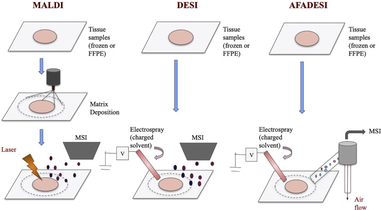
Metabolic heterogeneity of cancer contributes significantly to its poor treatment outcomes and prognosis. As a result, studies continue to focus on identifying new biomarkers and metabolic vulnerabilities, both of which depend on the understanding of altered metabolism in cancer. In the recent decades, the rise of mass spectrometry imaging (MSI) enables the detection of large numbers of small molecules in tissues. Therefore, researchers look to using MSI-mediated spatial metabolomics to further study the altered metabolites in cancer patients. In this review, we examined the two most commonly used spatial metabolomics techniques, MALDI-MSI and DESI-MSI, and some recent highlights of their applications in cancer studies. We also described AFADESI-MSI as a recent variation from the DESI-MSI and compare it with the two major techniques. Specifically, we discussed spatial metabolomics results in four types of heterogeneous malignancies, including breast cancer, esophageal cancer, glioblastoma and lung cancer. Multiple studies have effectively classified cancer tissue subtypes using altered metabolites information. In addition, distribution trends of key metabolites such as fatty acids, high-energy phosphate compounds, and antioxidants were identified. Therefore, while the visualization of finer distribution details requires further improvement of MSI techniques, past studies have suggested spatial metabolomics to be a promising direction to study the complexity of cancer pathophysiology.
癌症的代谢异质性是导致其治疗效果不佳和预后不良的重要原因。因此,相关研究持续聚焦于寻找新的生物标志物和代谢弱点,而这两者都依赖于对癌症中代谢改变的理解。近几十年来,质谱成像(MSI)技术的兴起使得在组织中检测大量小分子成为可能。因此,研究人员期望利用MSI介导的空间代谢组学进一步研究癌症患者体内改变的代谢物。在本综述中,我们考察了两种最常用的空间代谢组学技术,基质辅助激光解吸电离质谱成像(MALDI-MSI)和解吸电喷雾电离质谱成像(DESI-MSI),以及它们在癌症研究中的一些最新应用亮点。我们还介绍了作为DESI-MSI最新变体的常压敞开式解吸电喷雾电离质谱成像(AFADESI-MSI),并将其与这两种主要技术进行比较。具体而言,我们讨论了在四种异质性恶性肿瘤(包括乳腺癌、食管癌、胶质母细胞瘤和肺癌)中的空间代谢组学研究结果。多项研究已利用改变的代谢物信息有效地对癌症组织亚型进行了分类。此外还确定了脂肪酸、高能磷酸化合物和抗氧化剂等关键代谢物的分布趋势。因此,虽然更精细分布细节可视化需要MSI技术的进一步改进,但过去的研究表明空间代谢组学是研究癌症病理生理学复杂性的一个有前景的方向。