Institute of Infection and Global Health, University of Liverpoolgrid.10025.36, Liverpool, United Kingdom.
Faculty of Pharmacy, Mahidol University, Bangkok, Thailand.
mBio. 2022 Aug 30;13(4):e0102422. doi: 10.1128/mbio.01024-22. Epub 2022 Aug 4.
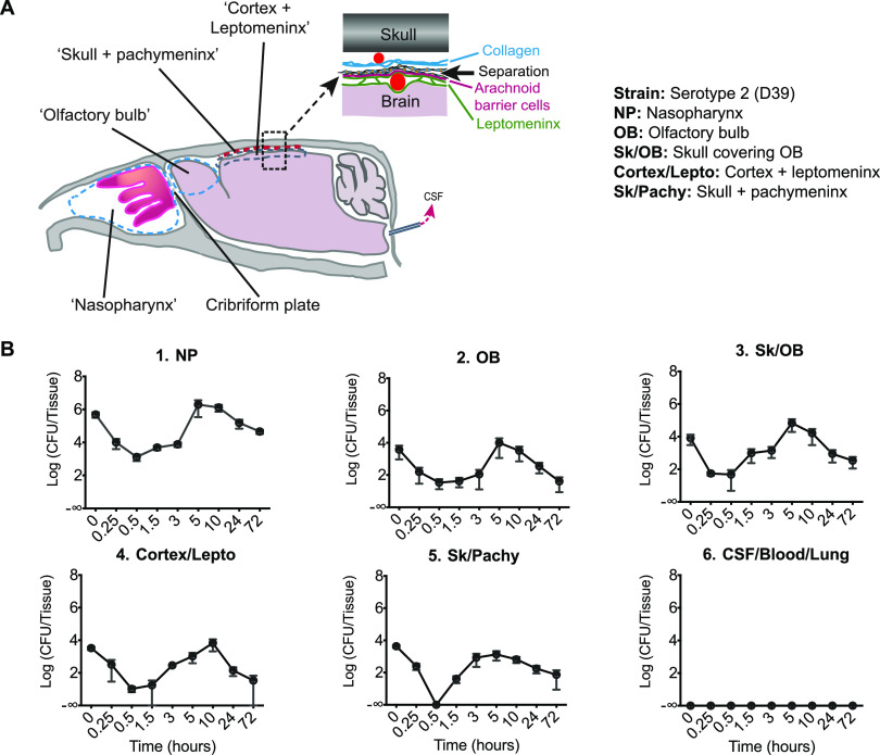
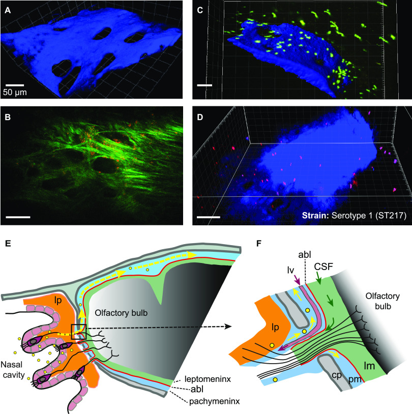
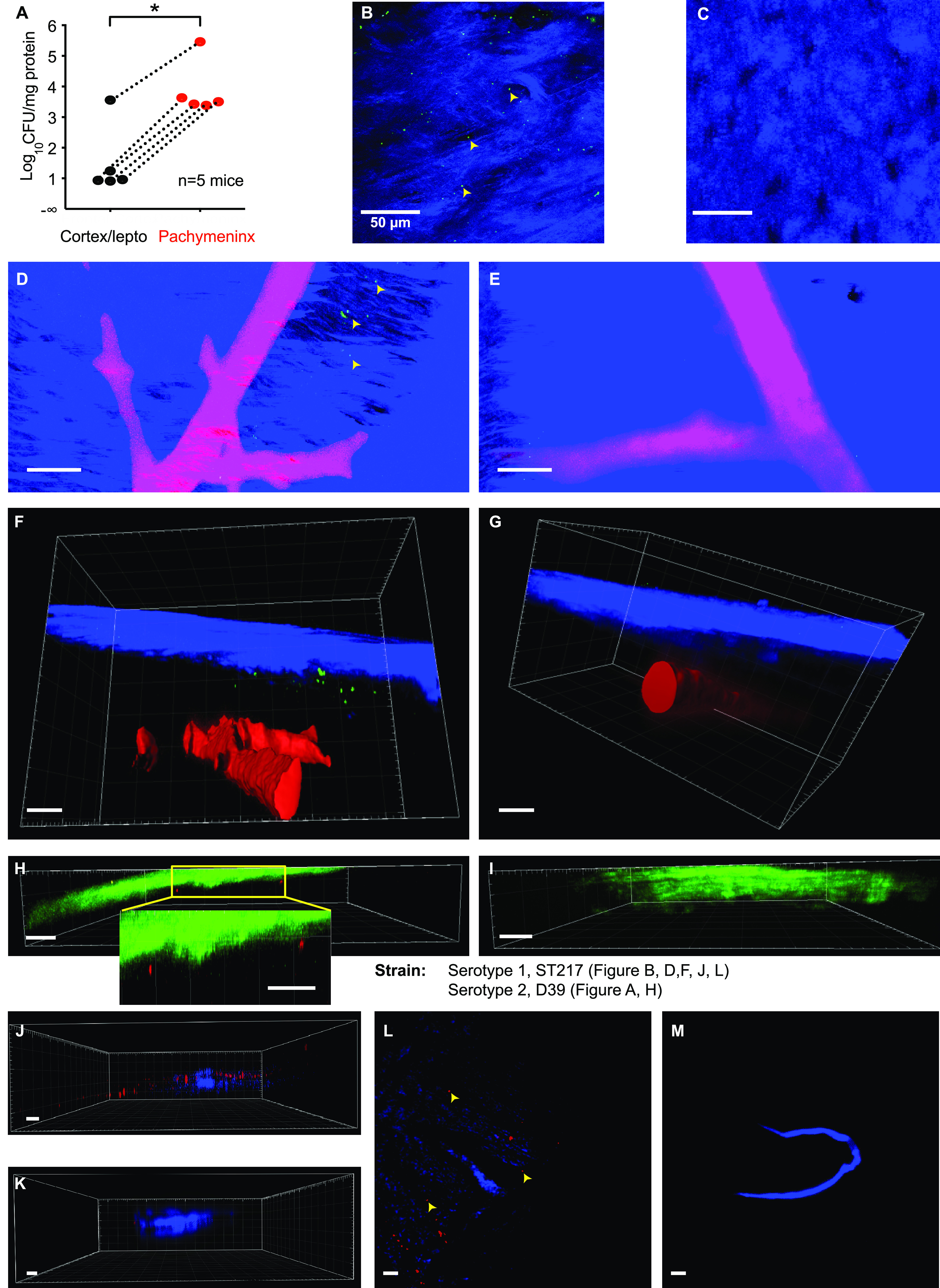
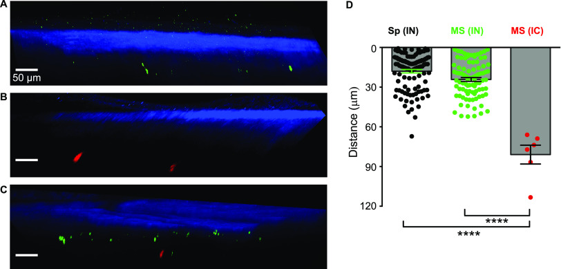
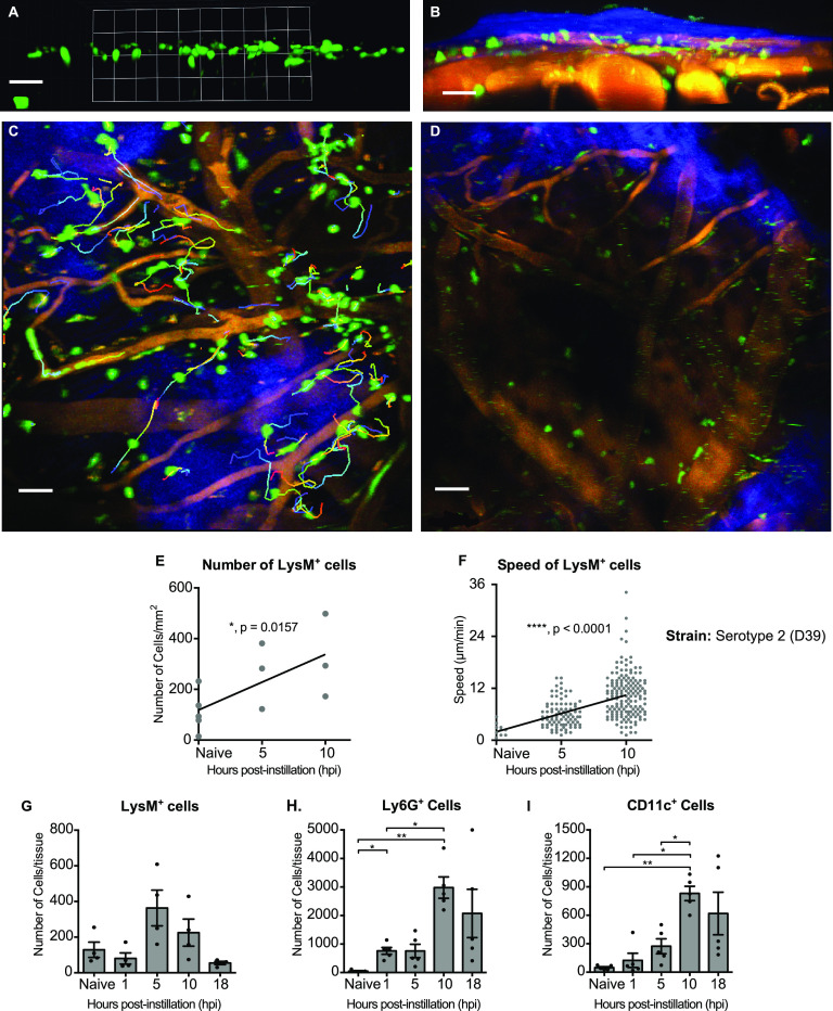
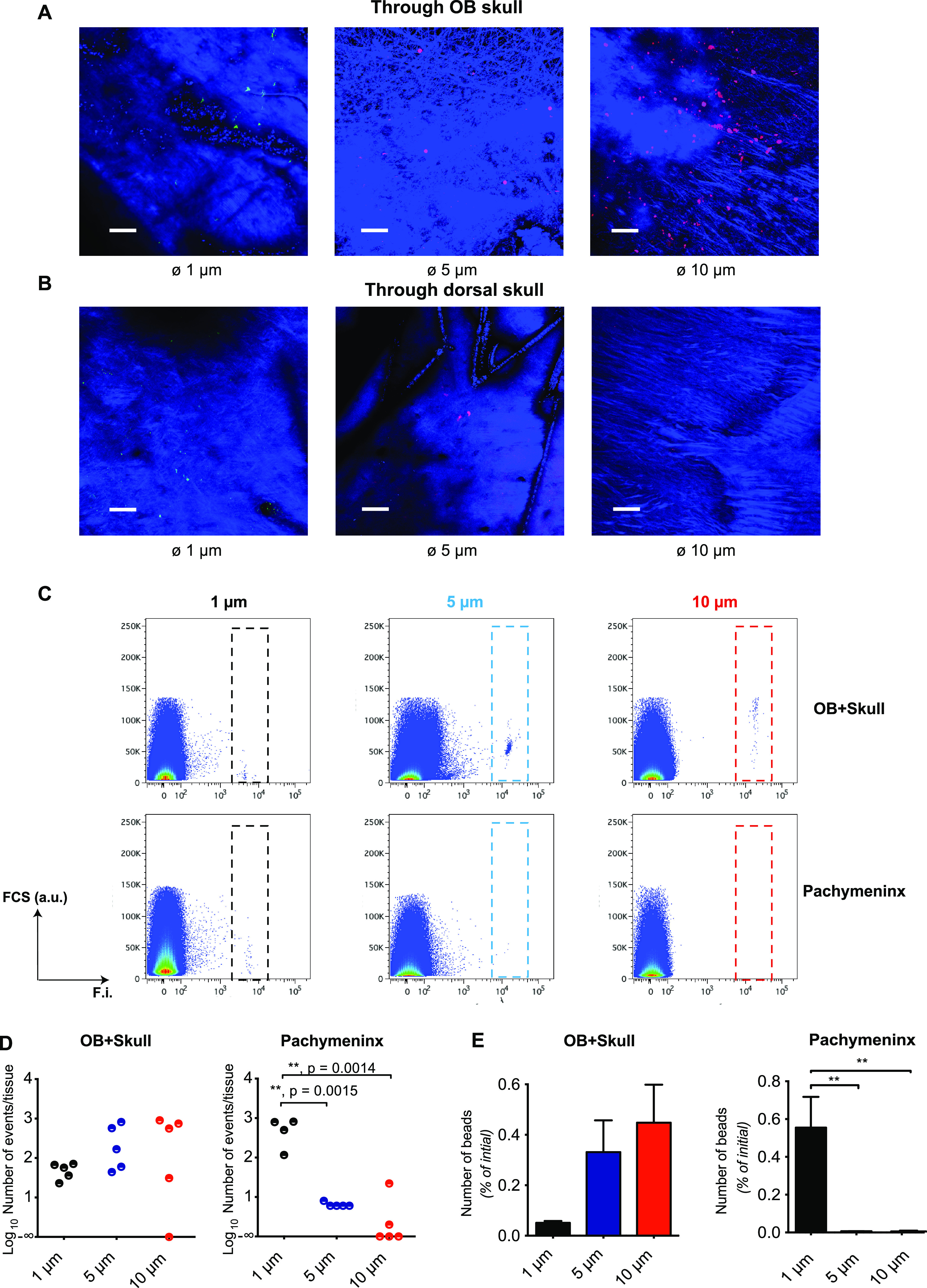
The entry routes and translocation mechanisms of microorganisms or particulate materials into the central nervous system remain obscure We report here that Streptococcus pneumoniae (pneumococcus), or polystyrene microspheres of similar size, appear in the meninges of the dorsal cortex of mice within minutes of inhaled delivery. Recovery of viable bacteria from dissected tissue and fluorescence microscopy show that up to at least 72 h, pneumococci and microspheres were predominantly found in the outer of the two meninges: the pachymeninx. No pneumococci were found in blood or cerebrospinal fluid. Intravital imaging through the skull, aligned with flow cytometry showed recruitment and activation of LysM cells in the dorsal pachymeninx at 5 and 10 hours following intranasal infection. Imaging of the cribriform plate suggested that both pneumococci and microspheres entered through the foramina via an inward flow of fluid connecting the nose to the pachymeninx. Our findings bring new insight into the varied mechanisms of pneumococcal invasion of the central nervous system, but they are also pertinent to the delivery of drugs to the brain and the entry of airborne particulate matter into the cranium. Using two-photon imaging, we show that pneumococci translocate from the nasopharynx to the dorsal meninges of a mouse in the absence of any bacteria found in blood or cerebrospinal fluid. Strikingly, this takes place within minutes of inhaled delivery of pneumococci, suggesting the existence of an inward flow of fluid connecting the nasopharynx to the meninges, rather than a receptor-mediated mechanism. We also show that this process is size dependent, as microspheres of the same size as pneumococci can translocate along the same pathway, while larger size microspheres cannot. Furthermore, we describe the host response to invasion of the outer meninges. Our study provides a completely new insight into the key initial events that occur during the translocation of pneumococci directly from the nasal cavity to the meninges, with relevance to the development of intranasal drug delivery systems and the investigations of brain damage caused by inhaled air pollutants.
微生物或颗粒物质进入中枢神经系统的进入途径和转移机制仍不清楚。我们在这里报告,肺炎链球菌(肺炎球菌)或类似大小的聚苯乙烯微球,在吸入后数分钟内出现在小鼠背侧皮质脑膜中。从解剖组织中回收活细菌和荧光显微镜观察表明,至少在 72 小时内,肺炎球菌和微球主要存在于两层脑膜的外层:硬脑膜。在血液或脑脊液中未发现肺炎球菌。通过颅骨进行的活体成像,与流式细胞术相结合,显示在鼻腔感染后 5 和 10 小时,背侧硬脑膜中的 LysM 细胞被募集和激活。对筛板的成像表明,肺炎球菌和微球都通过通过连接鼻子和硬脑膜的向内流动的液体进入小房孔。我们的发现为肺炎球菌侵入中枢神经系统的各种机制提供了新的见解,但它们也与将药物递送到大脑和空气中的颗粒物质进入颅骨有关。 使用双光子成像,我们表明肺炎球菌在没有任何血液或脑脊液中发现细菌的情况下从鼻咽转移到小鼠的背侧脑膜。引人注目的是,这发生在吸入肺炎球菌后数分钟内,表明存在将鼻咽连接到脑膜的向内流动,而不是受体介导的机制。我们还表明,该过程是大小依赖性的,因为与肺炎球菌大小相同的微球可以沿着相同的途径转移,而较大的微球则不能。此外,我们描述了宿主对脑膜外层入侵的反应。我们的研究为肺炎球菌从鼻腔直接转移到脑膜过程中发生的关键初始事件提供了全新的见解,这与鼻腔给药系统的发展以及对吸入空气污染物引起的脑损伤的研究有关。