Center for Brain Immunology and Glia (BIG), Washington University in St. Louis, St. Louis, MO 63110, USA; Department of Pathology and Immunology, School of Medicine, Washington University in St. Louis, St. Louis, MO 63110, USA.
Center for Brain Immunology and Glia (BIG), Washington University in St. Louis, St. Louis, MO 63110, USA; Department of Pathology and Immunology, School of Medicine, Washington University in St. Louis, St. Louis, MO 63110, USA.
Cell. 2021 Feb 18;184(4):1000-1016.e27. doi: 10.1016/j.cell.2020.12.040. Epub 2021 Jan 27.
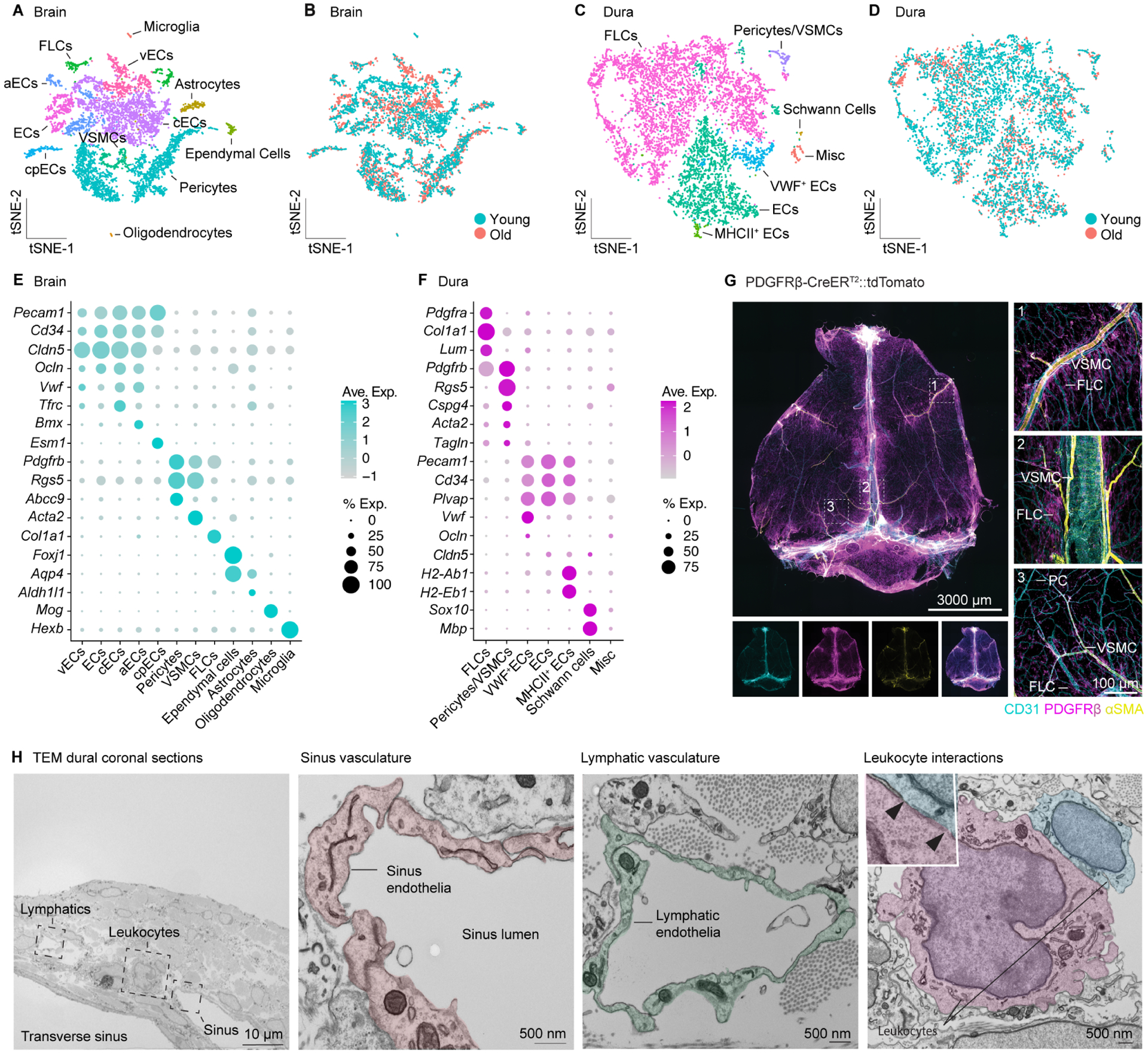
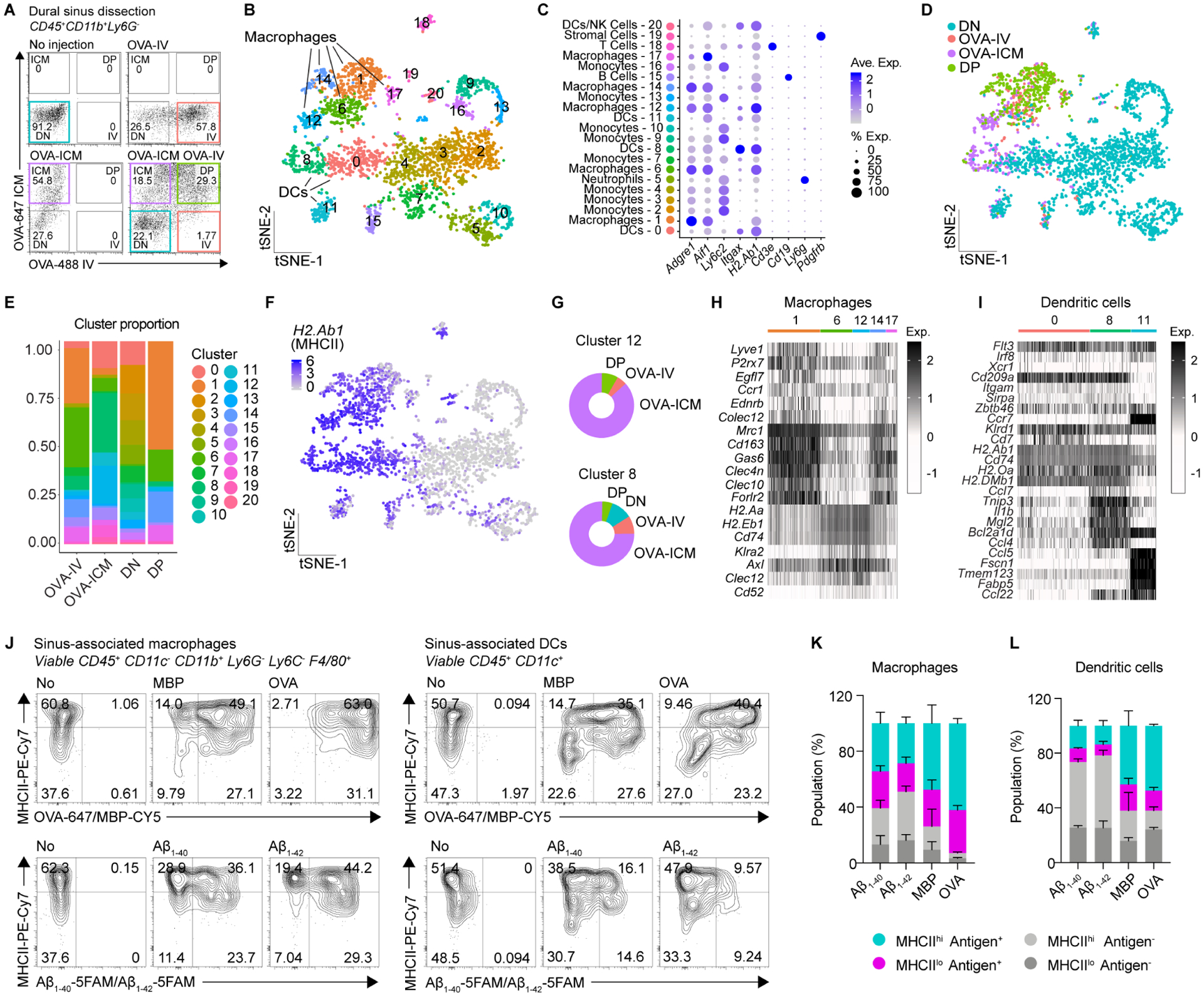
Despite the established dogma of central nervous system (CNS) immune privilege, neuroimmune interactions play an active role in diverse neurological disorders. However, the precise mechanisms underlying CNS immune surveillance remain elusive; particularly, the anatomical sites where peripheral adaptive immunity can sample CNS-derived antigens and the cellular and molecular mediators orchestrating this surveillance. Here, we demonstrate that CNS-derived antigens in the cerebrospinal fluid (CSF) accumulate around the dural sinuses, are captured by local antigen-presenting cells, and are presented to patrolling T cells. This surveillance is enabled by endothelial and mural cells forming the sinus stromal niche. T cell recognition of CSF-derived antigens at this site promoted tissue resident phenotypes and effector functions within the dural meninges. These findings highlight the critical role of dural sinuses as a neuroimmune interface, where brain antigens are surveyed under steady-state conditions, and shed light on age-related dysfunction and neuroinflammatory attack in animal models of multiple sclerosis.
尽管中枢神经系统 (CNS) 免疫特权的既定教条已经确立,但神经免疫相互作用在多种神经疾病中起着积极作用。然而,CNS 免疫监视的确切机制仍难以捉摸;特别是外周适应性免疫可以取样 CNS 来源抗原的解剖部位以及协调这种监视的细胞和分子介质。在这里,我们证明 CSF 中 CNS 来源的抗原在硬脑膜窦周围积聚,被局部抗原呈递细胞捕获,并呈递给巡逻 T 细胞。这种监视是由形成窦基质龛的内皮细胞和壁细胞实现的。T 细胞在该部位识别 CSF 来源的抗原,促进硬脑膜中的组织驻留表型和效应功能。这些发现强调了硬脑膜窦作为神经免疫界面的关键作用,在稳态条件下对脑抗原进行检测,并阐明了多发性硬化症动物模型中与年龄相关的功能障碍和神经炎症攻击。