Department of Neurology, Wake Forest Baptist Medical Center, Winston-Salem, North Carolina, USA.
Department of Neurology, Wake Forest School of Medicine, Winston-Salem, NC, USA.
J Neuroimaging. 2022 Nov;32(6):1013-1026. doi: 10.1111/jon.13031. Epub 2022 Aug 4.
Many studies have explored the possibility of using cranial ultrasound for discerning intracranial pathologies like tumors, hemorrhagic stroke, or subdural hemorrhage in clinical scenarios where computer tomography may not be accessible or feasible. The visualization of intracranial anatomy on B-mode ultrasound is challenging due to the presence of the skull that limits insonation to a few segments on the temporal bone that are thin enough to allow transcranial transmission of sound. Several artifacts are produced by hyperechoic signals inherent in brain and skull anatomy when images are created using temporal windows.
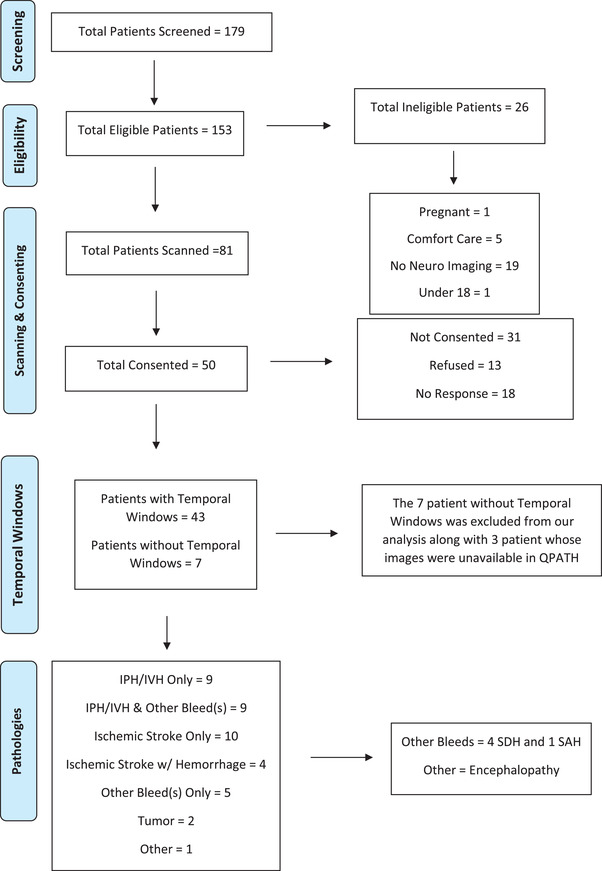
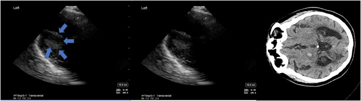
While the literature has investigated the accuracy of diagnosis of intracranial pathology with ultrasound, we lack a reference source for images acquired on cranial topography on B-mode ultrasound to illustrate the appearance of normal and abnormal structures of the brain and skull. Two investigators underwent hands-on training in Cranial point-of-care ultrasound (c-POCUS) and acquired multiple images from each patient to obtain the most in-depth images of brain to investigate all visible anatomical structures and pathology within 24 hours of any CT/MRI imaging done.
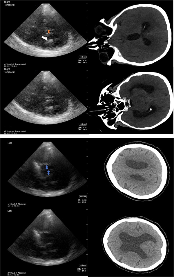
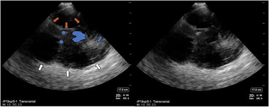
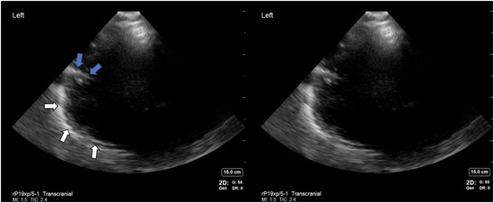
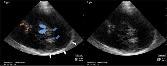
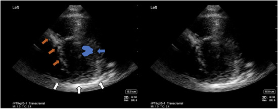
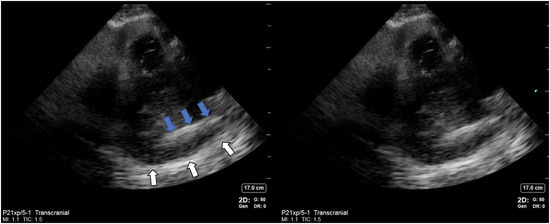
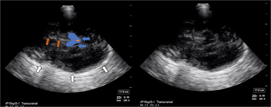
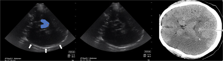
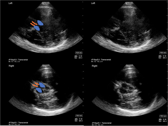
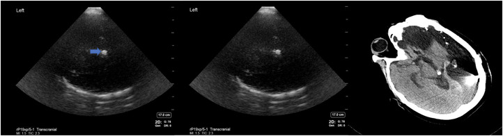
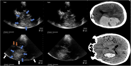
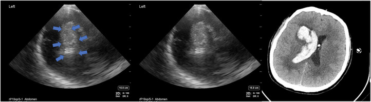
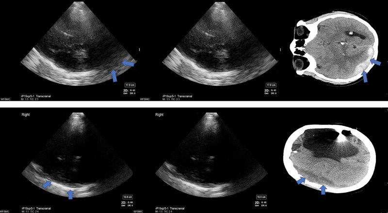
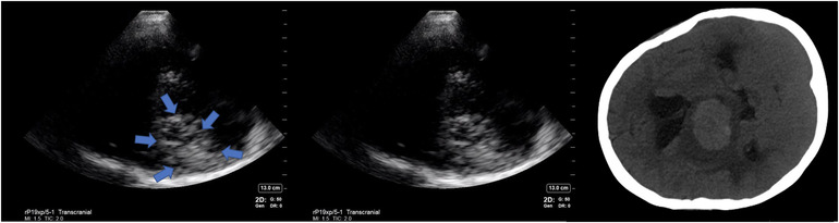
Most reproducible structures visible on c-POCUS included bony parts and parenchymal structures. Transcranial and abdominal presets were equivalent in elucidating anatomical structures. Brain pathology like parenchymal hemorrhage, cerebral edema, and hydrocephalus were also visualized.
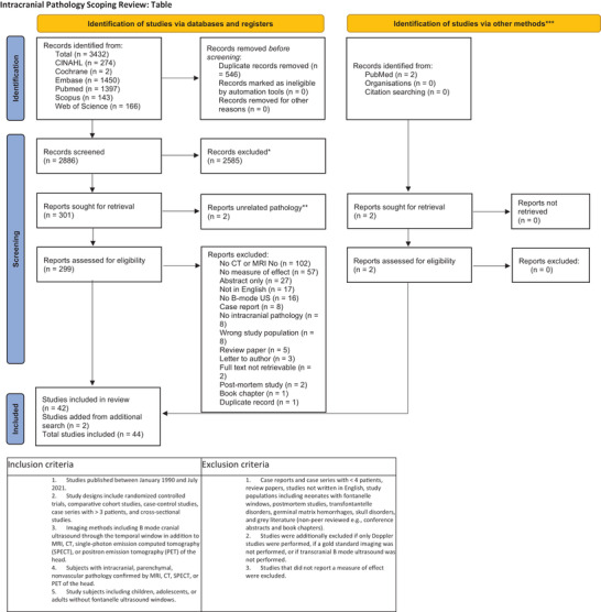
We present an illustrated anatomical atlas of cranial ultrasound B-mode images acquired in various pathologies in a critical care environment and compare our findings with published literature by performing a scoping review of literature on the subject.
许多研究探索了在计算机断层扫描(CT)不可用或不可行的临床情况下,使用颅超声识别颅内病变(如肿瘤、出血性中风或硬膜下血肿)的可能性。由于颅骨的存在,限制了对颞骨上几个足够薄以允许经颅声传输的骨段进行照射,因此 B 模式超声对颅内解剖结构的可视化具有挑战性。当使用颞窗创建图像时,脑和颅骨解剖结构固有的高回声信号会产生多种伪影。
虽然文献已经研究了超声诊断颅内病变的准确性,但我们缺乏关于颅骨 B 模式超声上颅顶采集的图像的参考资料,以说明脑和颅骨正常和异常结构的外观。两名调查员接受了颅急症超声(c-POCUS)的实践培训,并对每位患者进行了多次图像采集,以在任何 CT/MRI 成像后 24 小时内获得最深入的脑图像,以研究所有可见的解剖结构和病理学。
c-POCUS 上最可重复的可见结构包括骨结构和实质结构。经颅和腹部预设在阐明解剖结构方面是等效的。脑实质出血、脑水肿和脑积水等脑病理学也得到了可视化。
我们在重症监护环境中展示了各种病理情况下获得的颅超声 B 模式图像的解剖学图谱,并通过对该主题的文献进行范围综述,将我们的发现与已发表的文献进行比较。