University MS Center (UMSC) Hasselt - Pelt, Hasselt, Belgium.
SMRC Sports Medical Research Center, BIOMED Biomedical Research Institute, Faculty of Medicine and Life Sciences, Hasselt University, Hasselt, Belgium.
J Cachexia Sarcopenia Muscle. 2022 Oct;13(5):2537-2550. doi: 10.1002/jcsm.13050. Epub 2022 Aug 4.
Patients with multiple sclerosis (MS) experience reduced exercise tolerance that substantially reduces quality of life. The mechanisms underpinning exercise intolerance in MS are not fully clear. This study aimed to determine the contributions of the cardiopulmonary system and peripheral muscle in MS-induced exercise intolerance before and after exercise training.
Twenty-three patients with MS (13 women) and 20 age-matched and sex-matched healthy controls (13 women) performed a cardiopulmonary exercise test. Muscle fibre type composition, size, succinate dehydrogenase (SDH) activity, capillarity, and gene expression and proteins related to mitochondrial density were determined in vastus lateralis muscle biopsies. Nine MS patients (five women) were re-examined following a 12 week exercise training programme consisting of high-intensity cycling interval and resistance training.
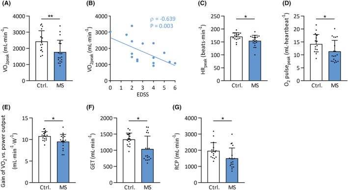
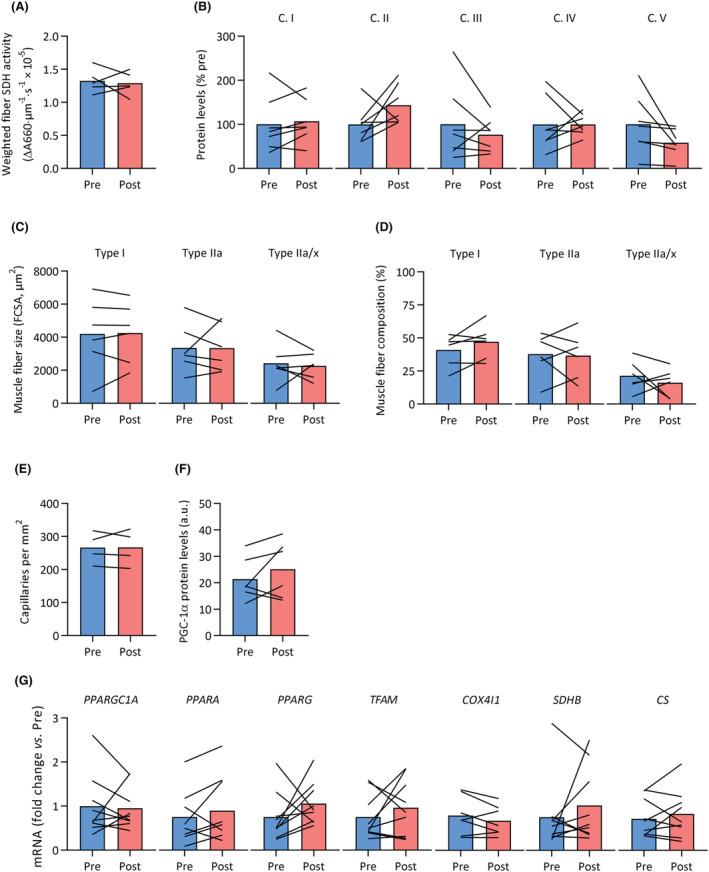
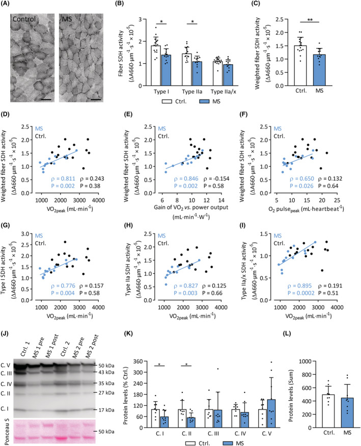
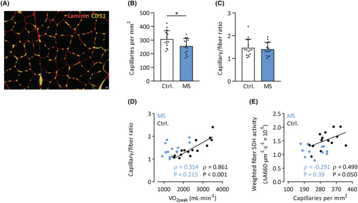
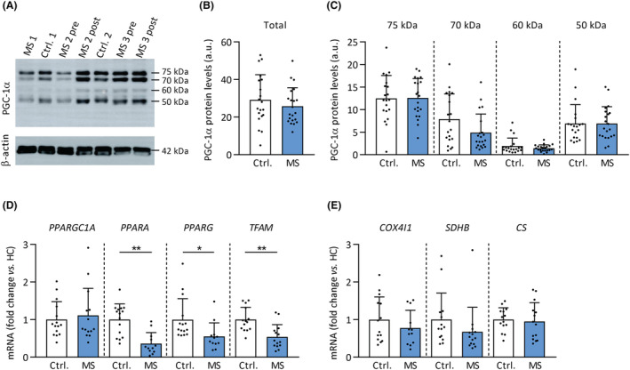
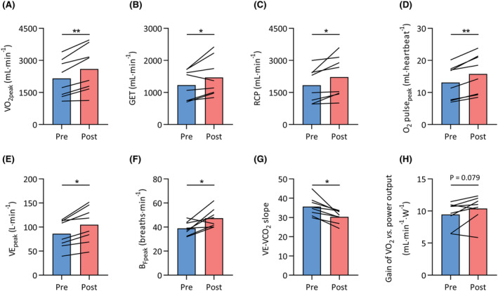
Patients with MS had lower maximal oxygen uptake compared with healthy controls (V̇O , 25.0 ± 8.5 vs. 35.7 ± 6.4 mL/kg/min, P < 0.001). The lower gas exchange threshold (MS: 14.5 ± 5.5 vs. controls: 19.7 ± 2.9 mL/kg/min, P = 0.01) and slope of V̇O versus work rate (MS: 9.5 ± 1.7 vs. controls: 10.8 ± 1.1 mL/min/W, P = 0.01) suggested an intramuscular contribution to exercise intolerance in patients with MS. Muscle SDH activity was 22% lower in MS (P = 0.004), and strongly correlated with several indices of whole-body exercise capacity in MS patients (e.g. V̇O , Spearman's ρ = 0.81, P = 0.002), but not healthy controls (ρ = 0.24, P = 0.38). In addition, protein levels of mitochondrial OXPHOS complexes I (-40%, P = 0.047) and II (-45%, P = 0.026) were lower in MS patients versus controls. Muscle capillary/fibre ratio correlated with V̇O in healthy controls (ρ = 0.86, P < 0.001) but not in MS (ρ = 0.35, P = 0.22), and did not differ between groups (1.41 ± 0.30 vs. 1.47 ± 0.38, P = 0.65). Expression of genes involved in mitochondrial function, such as PPARA, PPARG, and TFAM, was markedly reduced in muscle tissue samples of MS patients (all P < 0.05). No differences in muscle fibre type composition or size were observed between groups (all P > 0.05). V̇O increased by 23% following exercise training in MS (P < 0.001); however, no changes in muscle capillarity, SDH activity, gene or protein expression were observed (all P > 0.05).
Skeletal muscle oxidative phenotype (mitochondrial complex I and II content, SDH activity) is lower in patients with MS, contributing to reduced exercise tolerance. However, skeletal muscle mitochondria appeared resistant to the beneficial effects of exercise training, suggesting that other physiological systems, at least in part, drive the improvements in exercise capacity following exercise training in MS.
多发性硬化症(MS)患者的运动耐量降低,这大大降低了他们的生活质量。导致 MS 患者运动耐量降低的机制尚不完全清楚。本研究旨在确定心肺系统和外周肌肉在 MS 患者运动训练前后导致运动耐量降低的贡献。
23 名 MS 患者(13 名女性)和 20 名年龄和性别匹配的健康对照者(13 名女性)进行了心肺运动测试。在股外侧肌活检中测定了肌纤维类型组成、大小、琥珀酸脱氢酶(SDH)活性、毛细血管密度以及与线粒体密度相关的基因表达和蛋白。9 名 MS 患者(5 名女性)在完成 12 周的高强度循环间歇和阻力训练运动训练计划后再次接受检查。
与健康对照组相比,MS 患者的最大摄氧量(V̇O )较低(25.0 ± 8.5 比 35.7 ± 6.4 mL/kg/min,P < 0.001)。较低的气体交换阈值(MS:14.5 ± 5.5 比对照组:19.7 ± 2.9 mL/kg/min,P = 0.01)和 V̇O 与工作率的斜率(MS:9.5 ± 1.7 比对照组:10.8 ± 1.1 mL/min/W,P = 0.01)表明 MS 患者的肌肉内贡献导致运动耐量降低。MS 患者的肌肉 SDH 活性低 22%(P = 0.004),与 MS 患者的全身运动能力的多个指标强烈相关(例如,V̇O ,Spearman's ρ = 0.81,P = 0.002),但与健康对照组无关(ρ = 0.24,P = 0.38)。此外,线粒体 OXPHOS 复合物 I(-40%,P = 0.047)和 II(-45%,P = 0.026)的蛋白水平在 MS 患者中较对照组低。肌肉毛细血管/纤维比与健康对照组的 V̇O 相关(ρ = 0.86,P < 0.001),但与 MS 无关(ρ = 0.35,P = 0.22),且两组之间无差异(1.41 ± 0.30 比 1.47 ± 0.38,P = 0.65)。MS 患者的肌肉组织样本中,涉及线粒体功能的基因(如 PPARA、PPARG 和 TFAM)的表达明显降低(均 P < 0.05)。两组之间的肌纤维类型组成或大小无差异(均 P > 0.05)。MS 患者在运动训练后 V̇O 增加了 23%(P < 0.001);然而,肌肉毛细血管、SDH 活性、基因或蛋白表达没有变化(均 P > 0.05)。
MS 患者的骨骼肌氧化表型(线粒体复合物 I 和 II 含量、SDH 活性)较低,导致运动耐量降低。然而,骨骼肌线粒体对运动训练的有益作用似乎具有抗性,这表明其他生理系统至少部分地驱动了 MS 患者运动能力在运动训练后的改善。