Institute of Neuroscience, State Key Laboratory of Neuroscience, Center for Excellence in Brain Science and Intelligence Technology, Chinese Academy of Sciences, Shanghai, 200031, China.
University of Chinese Academy of Sciences, Beijing, 100049, China.
Neurosci Bull. 2022 Dec;38(12):1559-1568. doi: 10.1007/s12264-022-00923-9. Epub 2022 Aug 8.
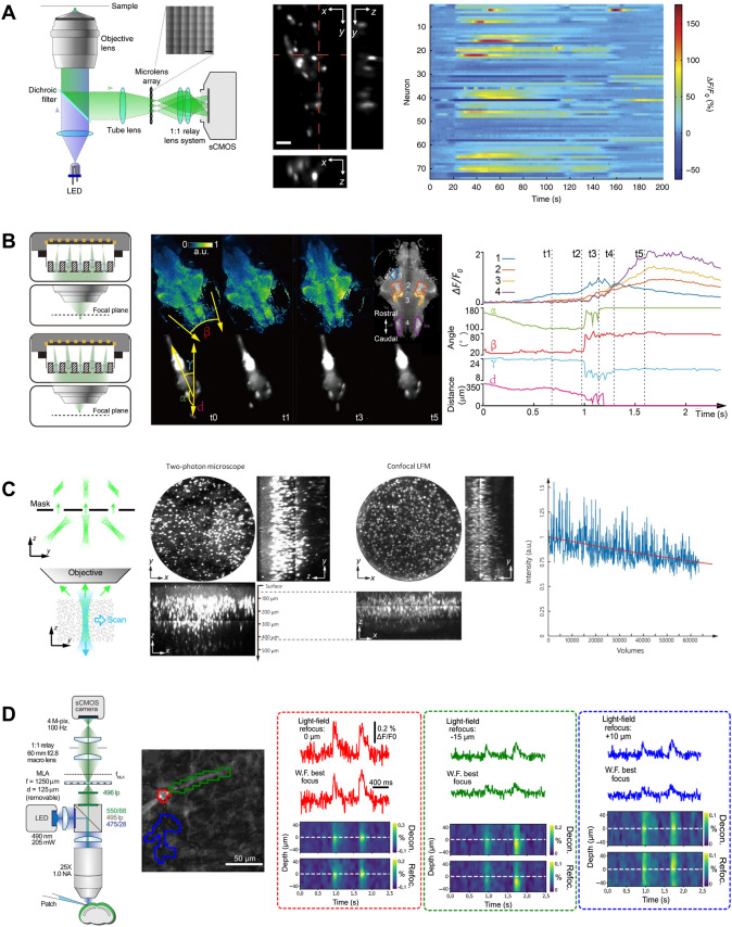
Recording the highly diverse and dynamic activities in large populations of neurons in behaving animals is crucial for a better understanding of how the brain works. To meet this challenge, extensive efforts have been devoted to developing functional fluorescent indicators and optical imaging techniques to optically monitor neural activity. Indeed, optical imaging potentially has extremely high throughput due to its non-invasive access to large brain regions and capability to sample neurons at high density, but the readout speed, such as the scanning speed in two-photon scanning microscopy, is often limited by various practical considerations. Among different imaging methods, light field microscopy features a highly parallelized 3D fluorescence imaging scheme and therefore promises a novel and faster strategy for functional imaging of neural activity. Here, we briefly review the working principles of various types of light field microscopes and their recent developments and applications in neuroscience studies. We also discuss strategies and considerations of optimizing light field microscopy for different experimental purposes, with illustrative examples in imaging zebrafish and mouse brains.
记录行为动物中大量神经元的高度多样化和动态活动对于更好地理解大脑如何工作至关重要。为了应对这一挑战,人们投入了大量精力来开发功能荧光指示剂和光学成像技术,以光学方式监测神经活动。事实上,由于光学成像可以非侵入性地访问大脑的大片区域,并以高密度对神经元进行采样,因此具有极高的通量潜力,但读出速度(例如双光子扫描显微镜中的扫描速度)通常受到各种实际考虑因素的限制。在不同的成像方法中,光场显微镜具有高度并行的 3D 荧光成像方案,因此有望成为神经活动功能成像的一种新颖而快速的策略。在这里,我们简要回顾了各种类型的光场显微镜的工作原理及其在神经科学研究中的最新发展和应用。我们还讨论了针对不同实验目的优化光场显微镜的策略和考虑因素,并提供了在成像斑马鱼和小鼠大脑方面的示例。