Department of Cardiology, Amsterdam UMC, Vrije Universiteit Amsterdam, Amsterdam Cardiovascular Sciences, De Boelelaan 1118, 1081 HV Amsterdam, The Netherlands.
Eur Heart J Cardiovasc Imaging. 2022 Aug 22;23(9):1182-1190. doi: 10.1093/ehjci/jeab245.
Various methods and post-processing software packages have been developed to quantify left atrial (LA) fibrosis using 3D late gadolinium-enhancement cardiac magnetic resonance (LGE-CMR) images. Currently, it remains unclear how the results of these methods and software packages interrelate.
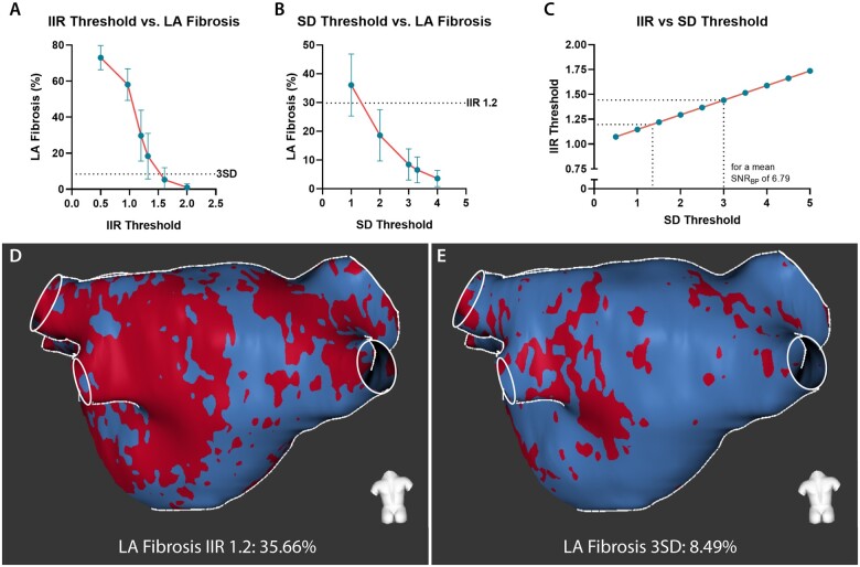
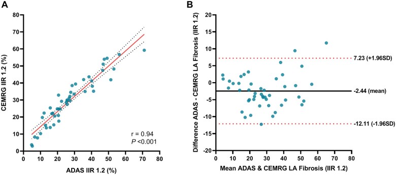
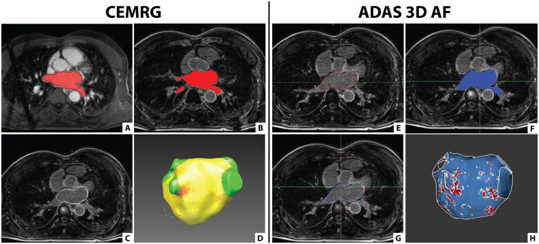
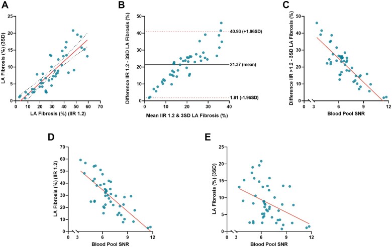
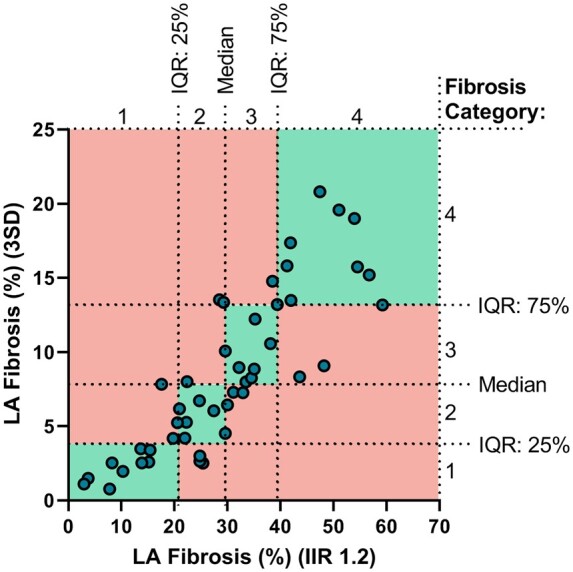
Forty-seven atrial fibrillation (AF) patients underwent 3D-LGE-CMR imaging prior to their AF ablation. LA fibrotic burden was derived from the images using open-source CEMRG software and commercially available ADAS 3D-LA software. Both packages were used to calculate fibrosis based on the image intensity ratio (IIR)-method. Additionally, CEMRG was used to quantify LA fibrosis using three standard deviations (3SD) above the mean blood pool signal intensity. Intraclass correlation coefficients were calculated to compare LA fibrosis quantification methods and different post-processing software outputs. The percentage of LA fibrosis assessed using IIR threshold 1.2 was significantly different from the 3SD-method (29.80 ± 14.15% vs. 8.43 ± 5.42%; P < 0.001). Correlation between the IIR-and SD-method was good (r = 0.85, P < 0.001) although agreement was poor [intraclass correlation coefficient (ICC) = 0.19; P < 0.001]. One-third of the patients were allocated to a different fibrosis category dependent on the used quantification method. Fibrosis assessment using CEMRG and ADAS 3D-LA showed good agreement for the IIR-method (ICC = 0.93; P < 0.001).
Both, the IIR1.2 and 3SD-method quantify atrial fibrotic burden based on atrial wall signal intensity differences. The discrepancy in the amount of LA fibrosis between these methods may have clinical implications when patients are classified according to their fibrotic burden. There was no difference in results between post-processing software packages to quantify LA fibrosis if an identical quantification method including the threshold was used.
目前已有多种方法和后处理软件包可用于通过三维晚期钆增强心脏磁共振(LGE-CMR)图像量化左心房(LA)纤维化。然而,目前尚不清楚这些方法和软件包的结果如何相互关联。
47 例心房颤动(AF)患者在 AF 消融前接受了 3D-LGE-CMR 成像。使用开源 CEMRG 软件和商业可用的 ADAS 3D-LA 软件从图像中得出 LA 纤维化负担。这两个软件包均基于图像强度比(IIR)方法计算纤维化。此外,CEMRG 还使用平均血池信号强度以上三个标准差(3SD)来量化 LA 纤维化。计算了组内相关系数以比较 LA 纤维化定量方法和不同后处理软件的输出。使用 IIR 阈值 1.2 评估的 LA 纤维化百分比与 3SD 方法明显不同(29.80±14.15%与 8.43±5.42%;P<0.001)。尽管一致性较差(组内相关系数(ICC)=0.19;P<0.001),但 IIR 与 SD 方法之间的相关性良好(r=0.85,P<0.001)。三分之一的患者根据使用的定量方法被分配到不同的纤维化类别。使用 CEMRG 和 ADAS 3D-LA 进行纤维化评估,对于 IIR 方法,两者具有良好的一致性(ICC=0.93;P<0.001)。
IIR1.2 和 3SD 方法均基于心房壁信号强度差异来量化心房纤维化负担。如果使用相同的包括阈值的定量方法,则这些方法之间 LA 纤维化量的差异可能会对根据纤维化负担对患者进行分类产生临床影响。如果使用相同的定量方法,包括阈值,则量化 LA 纤维化的后处理软件包之间的结果没有差异。