Wang Juan, Wang Le, Chen Xing, Liang Mao-Li, Wei Dong-Hui, Cao Wei, Zhang Jing
Department of Respiratory and Critical Care Medicine, Tianjin Medical University General Hospital, Tianjin 300052, China.
Iran J Basic Med Sci. 2022 Jun;25(6):755-761. doi: 10.22038/IJBMS.2022.64170.14133.
Cigarette smoke may play a direct role in proliferation of human pulmonary artery smooth muscle cells (HPASMCs). However, the mechanism involved and the effect of interventions remain unclear. We aimed to evaluate the effect of cigarette smoke extract (CSE) on HPASMCs, explore the role of inflammation and oxidative stress, and the effects of Tempol and PDTC in this process.
HPASMCs were subjected to normal control (NC), CSE, CSE+Tempol (CSE+T), and CSE+PDTC (CSE+P) groups. Proliferation of HPASMCs was measured by CCK-8 and Western blot. TNF-α, IL-6, MDA, and SOD levels were determined by ELISA and commercial kits. Nuclear translocation of NF-κB p65 was evaluated by western blot.
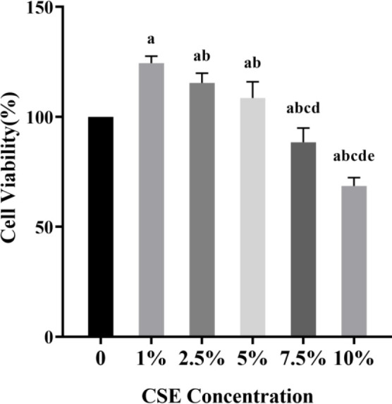
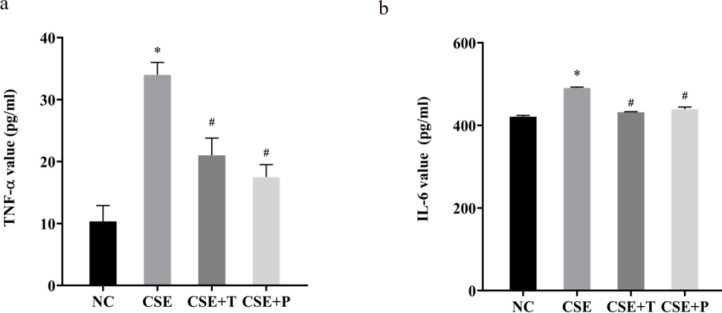
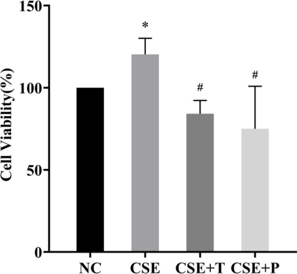
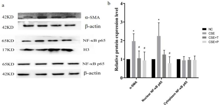
1%, 2.5%, and 5% CSE all promoted proliferation of HPASMCs, and effect of 1% CSE was the most significant, however, 7.5% and 10% CSE inhibited viability of cells (all <0.05). Compared with the NC group, TNF-α, IL-6, and MDA levels increased, SOD activity decreased (all <0.05), and NF-κB p65 expression in nuclei increased (=0.04) in the CSE group. Tempol and PDTC inhibited the proliferation of HPASMCs induced by CSE (all <0.05). And compared with the CSE group, TNF-α, IL-6, and MDA levels in CSE+T and CSE+P groups decreased, while SOD activity increased (all <0.05). Tempol reduced the expression of NF-κB p65 in nuclei but did not achieve a significant difference (=0.08). PDTC inhibited the nuclear translocation of NF-κB p65 (=0.03).
CSE stimulates HPASMCs proliferation in a certain concentration range. The CSE-induced proliferation of HPASMCs involved excessive inflammatory response and oxidative stress. Tempol and PDTC attenuate these effects of CSE on HPASMCs.
香烟烟雾可能在人肺动脉平滑肌细胞(HPASMCs)增殖中起直接作用。然而,其中涉及的机制以及干预措施的效果仍不清楚。我们旨在评估香烟烟雾提取物(CSE)对HPASMCs的影响,探讨炎症和氧化应激的作用,以及Tempol和PDTC在此过程中的作用。
将HPASMCs分为正常对照组(NC)、CSE组、CSE + Tempol组(CSE + T)和CSE + PDTC组(CSE + P)。通过CCK - 8和蛋白质免疫印迹法检测HPASMCs的增殖情况。采用酶联免疫吸附测定法(ELISA)和商用试剂盒测定肿瘤坏死因子-α(TNF-α)、白细胞介素-6(IL - 6)、丙二醛(MDA)和超氧化物歧化酶(SOD)水平。通过蛋白质免疫印迹法评估核因子-κB p65(NF-κB p65)的核转位情况。
1%、2.5%和5%的CSE均促进了HPASMCs的增殖,其中1% CSE的作用最为显著,然而,7.5%和10%的CSE抑制了细胞活力(均P<0.05)。与NC组相比,CSE组的TNF-α、IL - 6和MDA水平升高,SOD活性降低(均P<0.05),细胞核中NF-κB p65表达增加(P = 0.04)。Tempol和PDTC抑制了CSE诱导的HPASMCs增殖(均P<0.05)。并且与CSE组相比,CSE + T组和CSE + P组的TNF-α、IL - 6和MDA水平降低,而SOD活性升高(均P<0.05)。Tempol降低了细胞核中NF-κB p65的表达,但未达到显著差异(P = 0.08)。PDTC抑制了NF-κB p65的核转位(P = 0.03)。
CSE在一定浓度范围内刺激HPASMCs增殖。CSE诱导的HPASMCs增殖涉及过度的炎症反应和氧化应激。Tempol和PDTC减弱了CSE对HPASMCs的这些影响。