Yamasaki Seita, Miyake Makoto, Sakamoto Jiro, Tamura Akinori, Yamagami Shintaro, Nisiuchi Suguru, Yamane Keiichiro, Tamaki Yodo, Enomoto Soichiro, Kondo Hirokazu, Tamura Toshihiro
Department of Cardiology, Tenri Hospital, Tenri, Japan.
J Cardiol Cases. 2022 Apr 21;26(2):134-138. doi: 10.1016/j.jccase.2022.03.018. eCollection 2022 Aug.
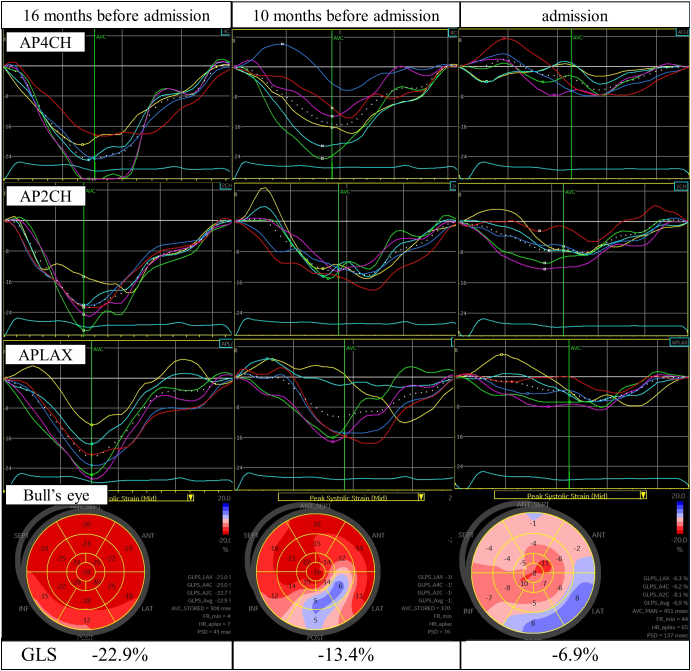
A 58-year-old man suffering from systemic sclerosis was admitted to our hospital because of heart failure. He developed atrioventricular block 4 months previously and had a pacemaker implanted, after which left ventricular wall motion markedly worsened. The global longitudinal strain was already decreased before the onset of atrioventricular block, although the left ventricular ejection fraction was normal. Right ventricular pacing was suspected to have caused overt left ventricular systolic dysfunction. Therefore, right ventricular pacing was upgraded to cardiac resynchronization therapy. After this change, the left ventricular ejection fraction improved to almost normal, but global longitudinal strain remained decreased. The findings in our case suggest that some patients with systemic sclerosis already have subclinical left ventricular systolic dysfunction before the onset of atrioventricular block. Additionally, right ventricular pacing may cause further deterioration of left ventricular systolic function and heart failure.
The possibility of subclinical left ventricular systolic dysfunction associated with systemic sclerosis should be considered when implanting a pacemaker. Speckle-tracking echocardiography may also be useful in the management of patients with systemic sclerosis.
一名58岁的系统性硬化症男性因心力衰竭入住我院。他在4个月前出现房室传导阻滞并植入了起搏器,此后左心室壁运动明显恶化。尽管左心室射血分数正常,但在房室传导阻滞发作前整体纵向应变就已降低。怀疑右心室起搏导致了明显的左心室收缩功能障碍。因此,将右心室起搏升级为心脏再同步治疗。这一改变后,左心室射血分数改善至几乎正常,但整体纵向应变仍降低。我们病例的结果表明,一些系统性硬化症患者在房室传导阻滞发作前就已经存在亚临床左心室收缩功能障碍。此外,右心室起搏可能会导致左心室收缩功能和心力衰竭进一步恶化。
植入起搏器时应考虑系统性硬化症相关亚临床左心室收缩功能障碍的可能性。斑点追踪超声心动图在系统性硬化症患者的管理中可能也有用。