• 文献检索
  • 文档翻译
  • 深度研究
  • 学术资讯
  • Suppr Zotero 插件Zotero 插件
  • 邀请有礼
  • 套餐&价格
  • 历史记录
应用&插件
Suppr Zotero 插件Zotero 插件浏览器插件Mac 客户端Windows 客户端微信小程序
定价
高级版会员购买积分包购买API积分包
服务
文献检索文档翻译深度研究API 文档MCP 服务
关于我们
关于 Suppr公司介绍联系我们用户协议隐私条款
关注我们

Suppr 超能文献

核心技术专利:CN118964589B侵权必究
粤ICP备2023148730 号-1Suppr @ 2026

文献检索

告别复杂PubMed语法,用中文像聊天一样搜索,搜遍4000万医学文献。AI智能推荐,让科研检索更轻松。

立即免费搜索

文件翻译

保留排版,准确专业,支持PDF/Word/PPT等文件格式,支持 12+语言互译。

免费翻译文档

深度研究

AI帮你快速写综述,25分钟生成高质量综述,智能提取关键信息,辅助科研写作。

立即免费体验

采用微流控 3D 发泡技术和动态培养的高度组织化多孔明胶支架用于软骨组织工程。

Highly Organized Porous Gelatin-Based Scaffold by Microfluidic 3D-Foaming Technology and Dynamic Culture for Cartilage Tissue Engineering.

机构信息

Department of Life Science, Fu Jen Catholic University, New Taipei City 242062, Taiwan.

Graduate Institute of Applied Science and Engineering, Fu Jen Catholic University, New Taipei City 242062, Taiwan.

出版信息

Int J Mol Sci. 2022 Jul 30;23(15):8449. doi: 10.3390/ijms23158449.

DOI:10.3390/ijms23158449
PMID:35955581
原文链接:https://pmc.ncbi.nlm.nih.gov/articles/PMC9369316/
Abstract

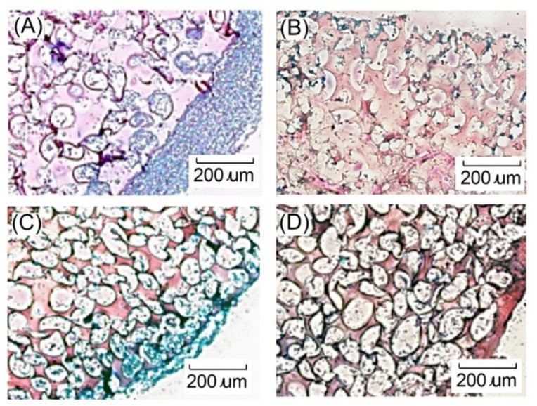
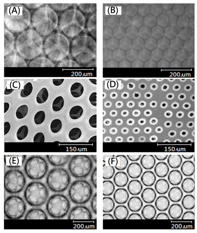
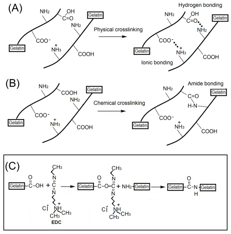
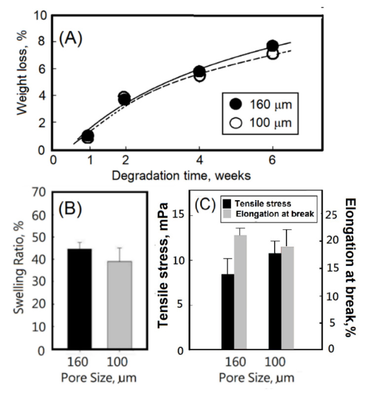
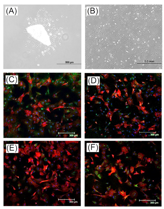
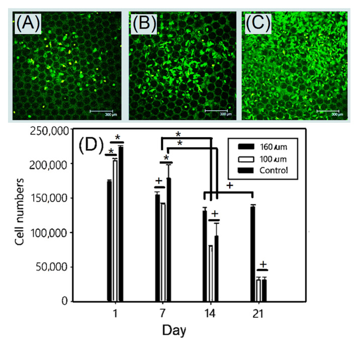
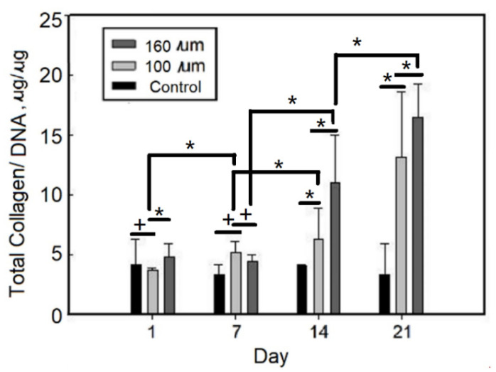
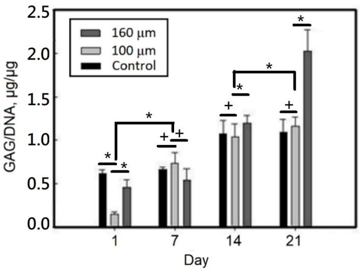
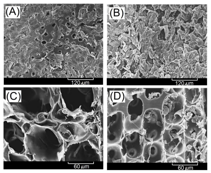
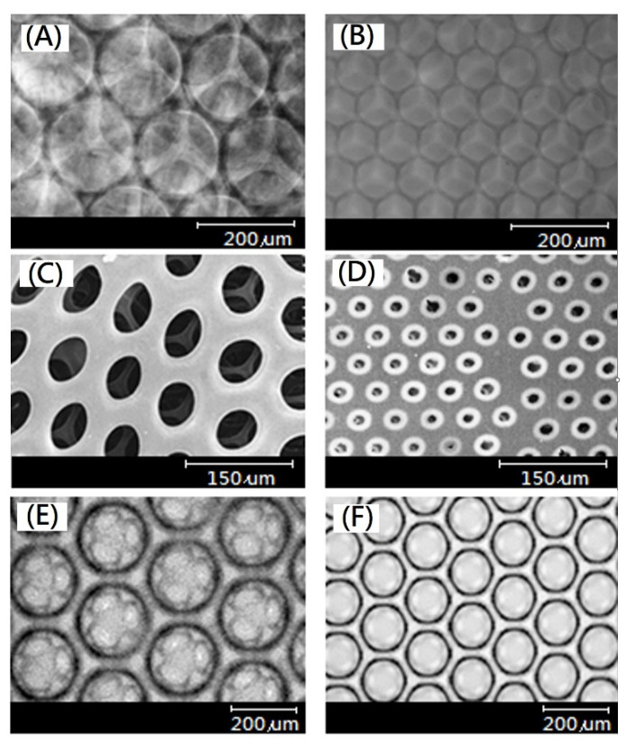
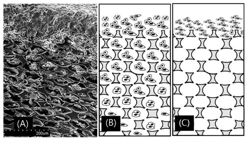
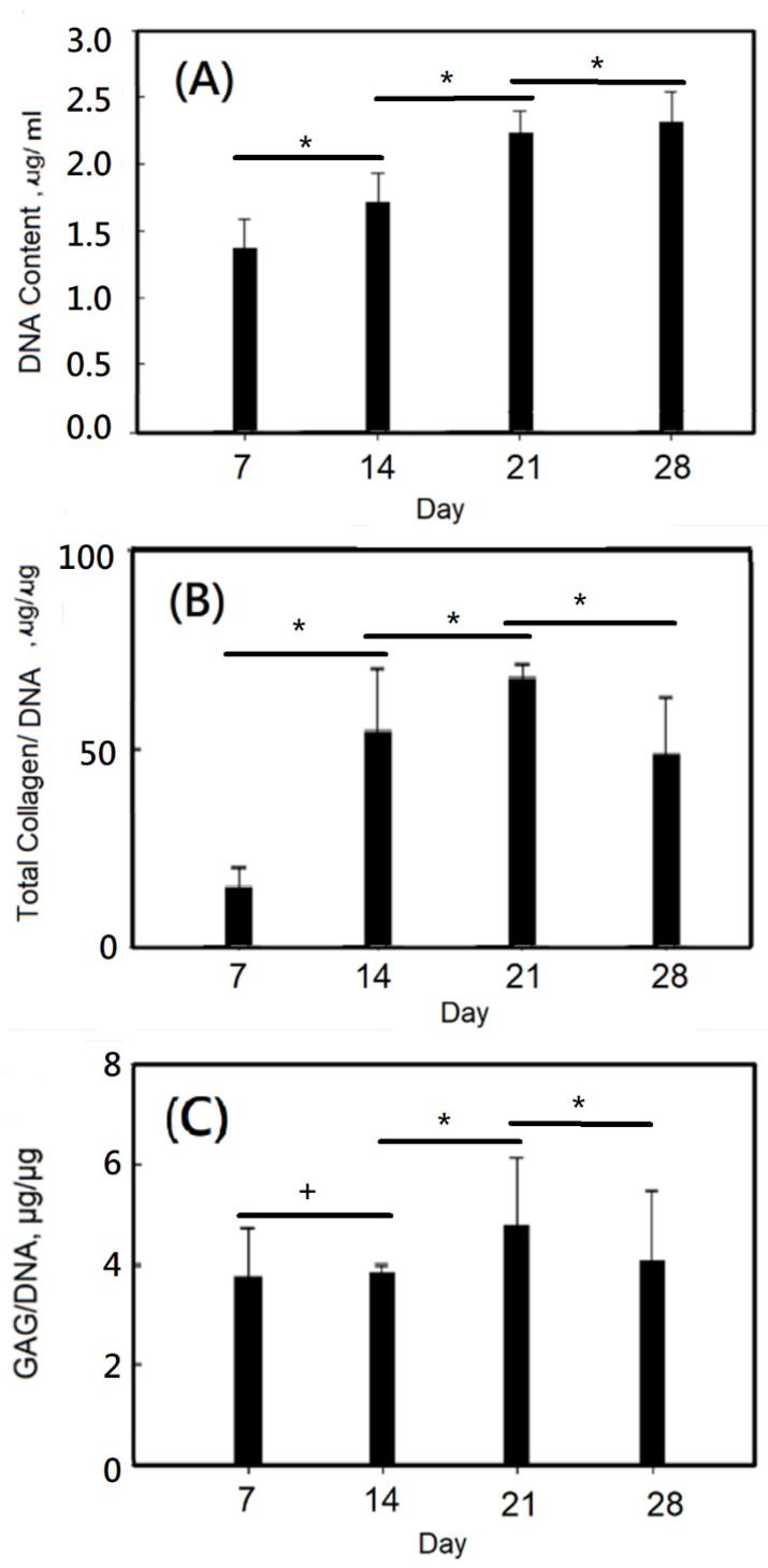
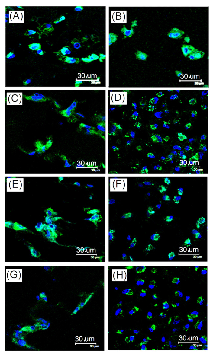
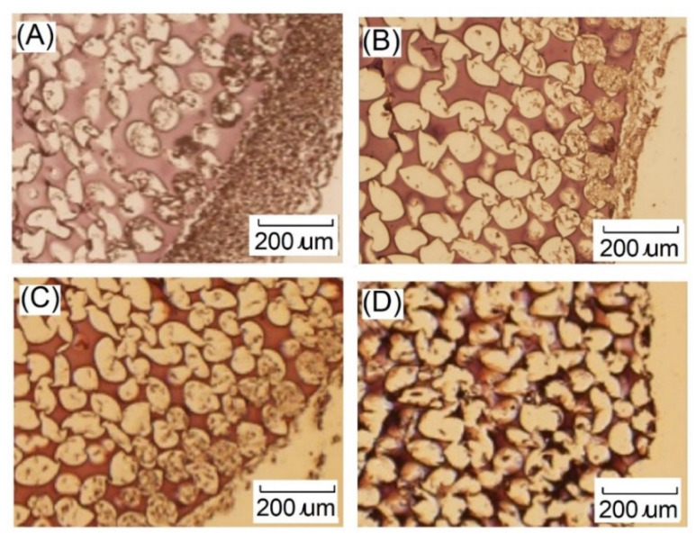
A gelatin-based hydrogel scaffold with highly uniform pore size and biocompatibility was fabricated for cartilage tissue engineering using microfluidic 3D-foaming technology. Mainly, bubbles with different diameters, such as 100 μm and 160 μm, were produced by introducing an optimized nitrogen gas and gelatin solution at an optimized flow rate, and N/gelatin bubbles were formed. Furthermore, a cross-linking agent (1-ethyl-3-(3-dimethyl aminopropyl)-carbodiimide, EDC) was employed for the cross-linking reaction of the gelatin-based hydrogel scaffold with uniform bubbles, and then the interface between the close cells were broken by degassing. The pore uniformity of the gelatin-based hydrogel scaffolds was confirmed by use of a bright field microscope, conjugate focus microscope and scanning electron microscope. The in vitro degradation rate, mechanical properties, and swelling rate of gelatin-based hydrogel scaffolds with highly uniform pore size were studied. Rabbit knee cartilage was cultured, and its extracellular matrix content was analyzed. Histological analysis and immunofluorescence staining were employed to confirm the activity of the rabbit knee chondrocytes. The chondrocytes were seeded into the resulting 3D porous gelatin-based hydrogel scaffolds. The growth conditions of the chondrocyte culture on the resulting 3D porous gelatin-based hydrogel scaffolds were evaluated by MTT analysis, live/dead cell activity analysis, and extracellular matrix content analysis. Additionally, a dynamic culture of cartilage tissue was performed, and the expression of cartilage-specific proteins within the culture time was studied by immunofluorescence staining analysis. The gelatin-based hydrogel scaffold encouraged chondrocyte proliferation, promoting the expression of collagen type II, aggrecan, and sox9 while retaining the structural stability and durability of the cartilage after dynamic compression and promoting cartilage repair.

摘要

采用微流控 3D 发泡技术,制备了具有高度均匀孔径和生物相容性的明胶基水凝胶支架用于软骨组织工程。主要是通过引入优化的氮气和明胶溶液以优化的流速来产生不同直径的气泡,如 100μm 和 160μm,形成 N/明胶气泡。此外,使用交联剂(1-乙基-3-(3-二甲基氨基丙基)碳二亚胺,EDC)对具有均匀气泡的明胶基水凝胶支架进行交联反应,然后通过脱气破坏封闭细胞之间的界面。使用明场显微镜、共轭焦显微镜和扫描电子显微镜证实了明胶基水凝胶支架的孔径均匀性。研究了具有高度均匀孔径的明胶基水凝胶支架的体外降解率、力学性能和溶胀率。培养兔膝关节软骨,并分析其细胞外基质含量。进行组织学分析和免疫荧光染色以确认兔膝关节软骨细胞的活性。将软骨细胞接种到所得的 3D 多孔明胶基水凝胶支架中。通过 MTT 分析、活/死细胞活性分析和细胞外基质含量分析评估软骨细胞在所得 3D 多孔明胶基水凝胶支架上的培养条件。此外,进行了软骨组织的动态培养,并通过免疫荧光染色分析研究了培养时间内软骨特异性蛋白的表达。明胶基水凝胶支架促进了软骨细胞的增殖,促进了 II 型胶原、聚集蛋白聚糖和 sox9 的表达,同时保留了软骨在动态压缩后的结构稳定性和耐久性,并促进了软骨修复。

https://cdn.ncbi.nlm.nih.gov/pmc/blobs/c876/9369316/a77277ebafb3/ijms-23-08449-g015.jpg
https://cdn.ncbi.nlm.nih.gov/pmc/blobs/c876/9369316/4690470f1e1e/ijms-23-08449-g001.jpg
https://cdn.ncbi.nlm.nih.gov/pmc/blobs/c876/9369316/6c9a5220b2a7/ijms-23-08449-g002.jpg
https://cdn.ncbi.nlm.nih.gov/pmc/blobs/c876/9369316/7c8d3393b5bf/ijms-23-08449-g003.jpg
https://cdn.ncbi.nlm.nih.gov/pmc/blobs/c876/9369316/c22f7f8335b0/ijms-23-08449-g004.jpg
https://cdn.ncbi.nlm.nih.gov/pmc/blobs/c876/9369316/0771c6951639/ijms-23-08449-g005.jpg
https://cdn.ncbi.nlm.nih.gov/pmc/blobs/c876/9369316/745cc76ae88c/ijms-23-08449-g006.jpg
https://cdn.ncbi.nlm.nih.gov/pmc/blobs/c876/9369316/1ca18b4849d2/ijms-23-08449-g007.jpg
https://cdn.ncbi.nlm.nih.gov/pmc/blobs/c876/9369316/d2af45462e1b/ijms-23-08449-g008.jpg
https://cdn.ncbi.nlm.nih.gov/pmc/blobs/c876/9369316/bceef8501f17/ijms-23-08449-g009.jpg
https://cdn.ncbi.nlm.nih.gov/pmc/blobs/c876/9369316/94a63300fd39/ijms-23-08449-g010.jpg
https://cdn.ncbi.nlm.nih.gov/pmc/blobs/c876/9369316/0136c4691610/ijms-23-08449-g011.jpg
https://cdn.ncbi.nlm.nih.gov/pmc/blobs/c876/9369316/1bdf1ccafb25/ijms-23-08449-g012.jpg
https://cdn.ncbi.nlm.nih.gov/pmc/blobs/c876/9369316/a0fde04b5c5e/ijms-23-08449-g013.jpg
https://cdn.ncbi.nlm.nih.gov/pmc/blobs/c876/9369316/77096322b7d7/ijms-23-08449-g014.jpg
https://cdn.ncbi.nlm.nih.gov/pmc/blobs/c876/9369316/a77277ebafb3/ijms-23-08449-g015.jpg

相似文献

1
Highly Organized Porous Gelatin-Based Scaffold by Microfluidic 3D-Foaming Technology and Dynamic Culture for Cartilage Tissue Engineering.采用微流控 3D 发泡技术和动态培养的高度组织化多孔明胶支架用于软骨组织工程。
Int J Mol Sci. 2022 Jul 30;23(15):8449. doi: 10.3390/ijms23158449.
2
[Research of diclofenac sodium-loaded gelatin scaffold with anti-inflammatory activity for promoting cartilage regeneration].[载双氯芬酸钠的具有抗炎活性的明胶支架促进软骨再生的研究]
Zhongguo Xiu Fu Chong Jian Wai Ke Za Zhi. 2023 Jan 15;37(1):91-100. doi: 10.7507/1002-1892.202207114.
3
Synthesis and in vitro evaluation of thermosensitive hydrogel scaffolds based on (PNIPAAm-PCL-PEG-PCL-PNIPAAm)/Gelatin and (PCL-PEG-PCL)/Gelatin for use in cartilage tissue engineering.基于(PNIPAAm-PCL-PEG-PCL-PNIPAAm)/明胶和(PCL-PEG-PCL)/明胶的温敏水凝胶支架的合成及体外评价及其在软骨组织工程中的应用。
J Biomater Sci Polym Ed. 2018 Jul;29(10):1185-1206. doi: 10.1080/09205063.2018.1447627. Epub 2018 Mar 5.
4
Three-dimensional dynamic fabrication of engineered cartilage based on chitosan/gelatin hybrid hydrogel scaffold in a spinner flask with a special designed steel frame.基于壳聚糖/明胶杂化水凝胶支架在带有特殊设计的钢框架的旋转瓶中三维动态构建工程化软骨。
Mater Sci Eng C Mater Biol Appl. 2015 Oct;55:384-92. doi: 10.1016/j.msec.2015.05.062. Epub 2015 May 27.
5
Fabrication of photo-crosslinked chitosan- gelatin scaffold in sodium alginate hydrogel for chondrocyte culture.用于软骨细胞培养的藻酸钠水凝胶中光交联壳聚糖-明胶支架的制备
Biomed Mater Eng. 2014;24(1):633-41. doi: 10.3233/BME-130851.
6
Dual Function of Glucosamine in Gelatin/Hyaluronic Acid Cryogel to Modulate Scaffold Mechanical Properties and to Maintain Chondrogenic Phenotype for Cartilage Tissue Engineering.氨基葡萄糖在明胶/透明质酸冷冻凝胶中的双重作用,用于调节支架力学性能并维持软骨组织工程中的软骨生成表型。
Int J Mol Sci. 2016 Nov 23;17(11):1957. doi: 10.3390/ijms17111957.
7
Laser sintered porous polycaprolacone scaffolds loaded with hyaluronic acid and gelatin-grafted thermoresponsive hydrogel for cartilage tissue engineering.负载透明质酸和明胶接枝温敏水凝胶的激光烧结多孔聚己内酯支架用于软骨组织工程
Biomed Mater Eng. 2013;23(6):533-43. doi: 10.3233/BME-130767.
8
In vitro expression of cartilage-specific markers by chondrocytes on a biocompatible hydrogel: implications for engineering cartilage tissue.软骨细胞在生物相容性水凝胶上对软骨特异性标志物的体外表达:对软骨组织工程的意义
Cell Transplant. 2001;10(8):755-63.
9
A highly organized three-dimensional alginate scaffold for cartilage tissue engineering prepared by microfluidic technology.采用微流控技术制备的高度有序的三维藻酸盐软骨组织工程支架。
Biomaterials. 2011 Oct;32(29):7118-26. doi: 10.1016/j.biomaterials.2011.06.018. Epub 2011 Jul 2.
10
3D Printed Chitosan Composite Scaffold for Chondrocytes Differentiation.3D 打印壳聚糖复合支架促进软骨细胞分化。
Curr Med Imaging. 2021;17(7):832-842. doi: 10.2174/1573405616666201217112939.

引用本文的文献

1
Antibacterial Crosslinker for Ternary PCL-Reinforced Hydrogels Based on Chitosan, Polyvinyl Alcohol, and Gelatin for Tissue Engineering.用于基于壳聚糖、聚乙烯醇和明胶的三元聚己内酯增强水凝胶的抗菌交联剂,用于组织工程
Polymers (Basel). 2025 May 29;17(11):1520. doi: 10.3390/polym17111520.
2
Cartilage repair: unleashing PRP's potential in organoid models.软骨修复:释放富血小板血浆在类器官模型中的潜力。
Cytotechnology. 2025 Jun;77(3):86. doi: 10.1007/s10616-025-00739-1. Epub 2025 Apr 3.
3
Hemocompatible gelatin-glycidyl methacrylate/graphene oxide composite hydrogels for vascular catheter applications.

本文引用的文献

1
Design and Characterization of a Bioinspired Polyvinyl Alcohol Matrix with Structural Foam-Wall Microarchitectures for Potential Tissue Engineering Applications.用于潜在组织工程应用的具有结构泡沫壁微结构的仿生聚乙烯醇基质的设计与表征
Polymers (Basel). 2022 Apr 13;14(8):1585. doi: 10.3390/polym14081585.
2
Evaluation and Preparation of a Designed Kartogenin Drug Delivery System (DDS) of Hydrazone-Linkage-Based pH Responsive mPEG-Hz--PCL Nanomicelles for Treatment of Osteoarthritis.基于腙键连接的pH响应性甲氧基聚乙二醇-腙-聚己内酯纳米胶束的设计型软骨生成素药物递送系统(DDS)用于骨关节炎治疗的评估与制备
Front Bioeng Biotechnol. 2022 Mar 9;10:816664. doi: 10.3389/fbioe.2022.816664. eCollection 2022.
3
用于血管导管应用的血液相容性明胶-甲基丙烯酸缩水甘油酯/氧化石墨烯复合水凝胶。
Sci Rep. 2025 Mar 25;15(1):10224. doi: 10.1038/s41598-025-93040-2.
4
Preparation of hydrogel microsphere and its application in articular cartilage injury.水凝胶微球的制备及其在关节软骨损伤中的应用。
Mater Today Bio. 2025 Mar 8;31:101641. doi: 10.1016/j.mtbio.2025.101641. eCollection 2025 Apr.
5
Gradient scaffolds in bone-soft tissue interface engineering: Structural characteristics, fabrication techniques, and emerging trends.骨-软组织界面工程中的梯度支架:结构特征、制造技术及新趋势
J Orthop Translat. 2025 Jan 28;50:333-353. doi: 10.1016/j.jot.2024.10.015. eCollection 2025 Jan.
6
Digital light processing printing of non-modified protein-only compositions.仅含未修饰蛋白质的组合物的数字光处理打印
Mater Today Bio. 2024 Dec 6;30:101384. doi: 10.1016/j.mtbio.2024.101384. eCollection 2025 Feb.
7
Dynamic-Cross-Linked, Regulated, and Controllable Mineralization Degree and Morphology of Collagen Biomineralization.胶原蛋白生物矿化的动态交联、调控及可控矿化程度与形态
J Funct Biomater. 2024 Nov 22;15(12):356. doi: 10.3390/jfb15120356.
8
Advancements in chondrocyte 3-dimensional embedded culture: Implications for tissue engineering and regenerative medicine.软骨细胞三维包埋培养的进展:对组织工程和再生医学的启示。
Biomed J. 2024 Sep 3;48(2):100786. doi: 10.1016/j.bj.2024.100786.
9
Osteoproductivity in Experimentally Induced Cranial Bone Defects in Rabbits.兔实验性颅骨骨缺损中的骨生成能力
Iran Biomed J. 2023 Sep 9;26(6):366-74. doi: 10.61186/ibj.3940.
10
Emerging Advances in Microfluidic Hydrogel Droplets for Tissue Engineering and STEM Cell Mechanobiology.用于组织工程和干细胞力学生物学的微流控水凝胶微滴的新进展
Gels. 2023 Oct 1;9(10):790. doi: 10.3390/gels9100790.
Characterization of Composite Nano-Bioscaffolds Based on Collagen and Supercritical Fluids-Assisted Decellularized Fibrous Extracellular Matrix.
基于胶原蛋白和超临界流体辅助去细胞化纤维细胞外基质的复合纳米生物支架的表征
Polymers (Basel). 2021 Dec 10;13(24):4326. doi: 10.3390/polym13244326.
4
Photocrosslinked gelatin hydrogel improves wound healing and skin flap survival by the sustained release of basic fibroblast growth factor.光交联明胶水凝胶通过持续释放碱性成纤维细胞生长因子促进伤口愈合和皮瓣存活。
Sci Rep. 2021 Nov 29;11(1):23094. doi: 10.1038/s41598-021-02589-1.
5
Characteristics and Preparation of Designed Alginate-Based Composite Scaffold Membranes with Decellularized Fibrous Micro-Scaffold Structures from Porcine Skin.具有来自猪皮的脱细胞纤维微支架结构的设计藻酸盐基复合支架膜的特性与制备
Polymers (Basel). 2021 Oct 9;13(20):3464. doi: 10.3390/polym13203464.
6
Biological evaluations of decellularized extracellular matrix collagen microparticles prepared based on plant enzymes and aqueous two-phase method.基于植物酶和双水相法制备的脱细胞细胞外基质胶原微粒的生物学评价
Regen Biomater. 2021 Mar 13;8(2):rbab002. doi: 10.1093/rb/rbab002. eCollection 2021 Mar.
7
Characterization of Gelatin Hydrogels Cross-Linked with Microbial Transglutaminase as Engineered Skeletal Muscle Substrates.用微生物转谷氨酰胺酶交联的明胶水凝胶作为工程化骨骼肌基质的表征
Bioengineering (Basel). 2021 Jan 6;8(1):6. doi: 10.3390/bioengineering8010006.
8
Tailoring the material properties of gelatin hydrogels by high energy electron irradiation.通过高能电子辐照定制明胶水凝胶的材料特性。
J Mater Chem B. 2014 Jul 21;2(27):4297-4309. doi: 10.1039/c4tb00429a. Epub 2014 Jun 6.
9
Multi-material 3D bioprinting of porous constructs for cartilage regeneration.多材料 3D 生物打印多孔结构用于软骨再生。
Mater Sci Eng C Mater Biol Appl. 2020 Apr;109:110578. doi: 10.1016/j.msec.2019.110578. Epub 2019 Dec 20.
10
Salinomycin causes dedifferentiation via the extracellular signal-regulated kinase (ERK) pathway in rabbit articular chondrocytes.沙利霉素通过细胞外信号调节激酶(ERK)途径导致兔关节软骨细胞去分化。
J Pharmacol Sci. 2015 Feb;127(2):196-202. doi: 10.1016/j.jphs.2014.12.009. Epub 2014 Dec 24.