Del Vecchio Sharon J, Urquhart Aaron J, Dong Xin, Ellis Robert J, Ng Keng Lim, Samaratunga Hemamali, Gustafson Sonja, Galloway Graham J, Gobe Glenda C, Wood Simon, Mountford Carolyn E
Kidney Disease Research Collaborative, Translational Research Institute, Princess Alexandra Hospital, The University of Queensland, Brisbane, Australia.
Department of Radiology, Princess Alexandra Hospital, Woolloongabba, Brisbane, Australia.
Transl Androl Urol. 2022 Jul;11(7):929-942. doi: 10.21037/tau-21-1082.
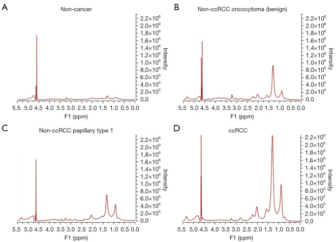
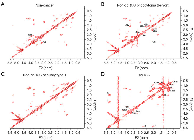
Routinely used clinical scanners, such as computed tomography (CT), magnetic resonance imaging (MRI) and ultrasound (US), are unable to distinguish between aggressive and indolent tumor subtypes in masses localized to the kidney, often leading to surgical overtreatment. The results of the current investigation demonstrate that chemical differences, detected in human kidney biopsies using two-dimensional COrrelated SpectroscopY (2D L-COSY) and evaluated using multivariate statistical analysis, can distinguish these subtypes.
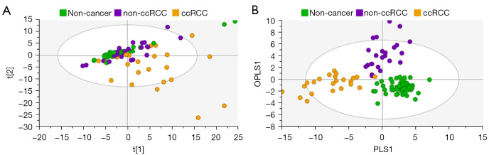
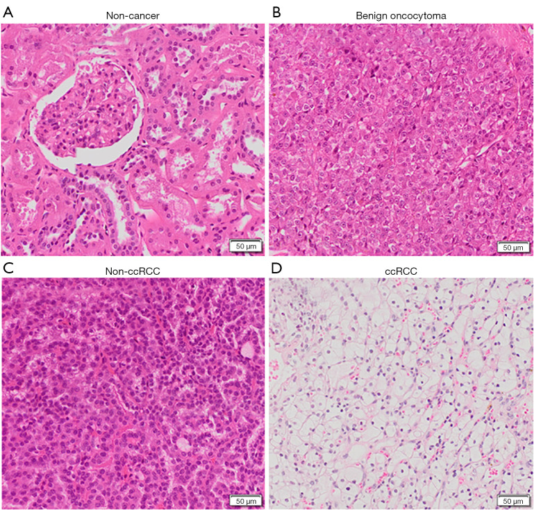
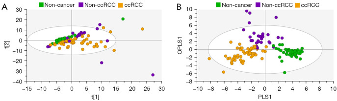
One hundred and twenty-six biopsy samples from patients with a confirmed enhancing kidney mass on abdominal imaging were analyzed as part of the training set. A further forty-three samples were used for model validation. In patients undergoing radical nephrectomy, biopsies of non-cancer kidney cortical tissue were also collected as a non-cancer control group. Spectroscopy data were analyzed using multivariate statistical analysis, including principal component analysis (PCA) and orthogonal projection to latent structures with discriminant analysis (OPLS-DA), to identify biomarkers in kidney cancer tissue that was also classified using the gold-standard of histopathology.
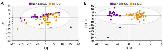
The data analysis methodology showed good separation between clear cell renal cell carcinoma (ccRCC) versus non-clear cell RCC (non-ccRCC) and non-cancer cortical tissue from the kidneys of tumor-bearing patients. Variable Importance for the Projection (VIP) values, and OPLS-DA loadings plots were used to identify chemical species that correlated significantly with the histopathological classification. Model validation resulted in the correct classification of 37/43 biopsy samples, which included the correct classification of 15/17 ccRCC biopsies, achieving an overall predictive accuracy of 86%, Those chemical markers with a VIP value >1.2 were further analyzed using univariate statistical analysis. A subgroup analysis of 47 tumor tissues arising from T1 tumors revealed distinct separation between ccRCC and non-ccRCC tissues.
This study provides metabolic insights that could have future diagnostic and/or clinical value. The results of this work demonstrate a clear separation between clear cell and non-ccRCC and non-cancer kidney tissue from tumor-bearing patients. The clinical translation of these results will now require the development of a one-dimensional (1D) magnetic resonance spectroscopy (MRS) protocol, for the kidney, using an clinical MRI scanner.
常规使用的临床扫描仪,如计算机断层扫描(CT)、磁共振成像(MRI)和超声(US),无法区分局限于肾脏的肿块中侵袭性和惰性肿瘤亚型,这常常导致手术过度治疗。当前研究结果表明,使用二维相关光谱法(2D L-COSY)在人肾活检中检测到并经多变量统计分析评估的化学差异能够区分这些亚型。
作为训练集的一部分,对126例经腹部成像确诊为肾脏强化肿块患者的活检样本进行了分析。另外43个样本用于模型验证。在接受根治性肾切除术的患者中,还收集了非癌性肾皮质组织活检样本作为非癌对照组。使用多变量统计分析,包括主成分分析(PCA)和带有判别分析的潜在结构正交投影(OPLS-DA),对光谱数据进行分析,以识别肾癌组织中的生物标志物,这些组织也使用组织病理学金标准进行了分类。
数据分析方法显示,透明细胞肾细胞癌(ccRCC)与非透明细胞肾细胞癌(non-ccRCC)以及荷瘤患者肾脏的非癌性皮质组织之间有良好的区分。使用投影变量重要性(VIP)值和OPLS-DA载荷图来识别与组织病理学分类显著相关的化学物质。模型验证结果为43个活检样本中的37个正确分类,其中包括17个ccRCC活检样本中的15个正确分类,总体预测准确率达到86%。对VIP值>1.2的那些化学标志物使用单变量统计分析进一步分析。对47个源于T1肿瘤的肿瘤组织进行的亚组分析显示,ccRCC和non-ccRCC组织之间有明显区分。
本研究提供了可能具有未来诊断和/或临床价值的代谢见解。这项工作的结果表明,透明细胞与non-ccRCC以及荷瘤患者的非癌性肾组织之间有明显区分。这些结果的临床转化现在需要开发一种使用临床MRI扫描仪对肾脏进行的一维(1D)磁共振波谱(MRS)方案。