Cerne John W, Liu Sophia, Umair Muhammad, Pathrose Ashitha, Moore Jackson E, Allen Bradley D, Markl Michael, Carr James C, Savas Hatice, Wilsbacher Lisa, Avery Ryan
Department of Radiology, Northwestern University Feinberg School of Medicine, 420 East Superior St, Chicago, IL, 60611, USA.
Biomedical Engineering, Northwestern University McCormick School of Engineering and Applied Science, Evanston, USA.
Eur J Hybrid Imaging. 2022 Aug 15;6(1):16. doi: 10.1186/s41824-022-00136-3.
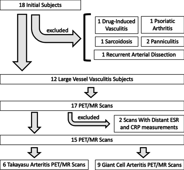
Large vessel vasculitis (LVV) can be characterized based on symptom severity, and this characterization helps clinicians decide upon treatment approach. Our aim was to compare the imaging findings of combined modality positron emission tomography/magnetic resonance (PET/MR) and inflammatory markers between severe and non-severe LVV. A retrospective query was performed to identify all patients with LVV who underwent PET/MR at our institution between January 2015 and January 2021.
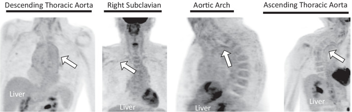
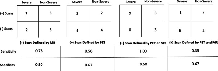
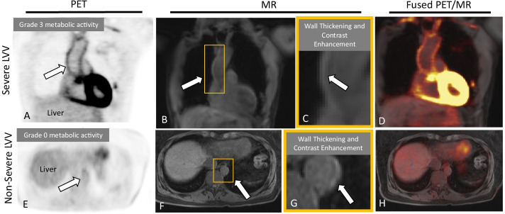
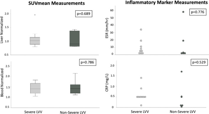
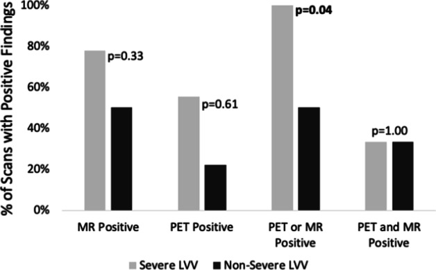
Eleven patients (nine females; age 62.2 ± 16.4 years) underwent 15 PET/MR scans. Positivity was defined by findings indicative of active LVV on each modality: PET positive if vessel metabolic activity > liver metabolic activity; MR positive if wall thickening or contrast enhancement. When positive PET or positive MR findings were considered a positive scan, LVV patients with severe disease (n = 9 scans) showed a higher number of positive scans (n = 9) compared to the number of positive scans in non-severe patients (n = 3) (p < 0.05). The sensitivity and specificity for the detection of severe LVV were 1.00 and 0.50, respectively. When only the presence of both positive PET and positive MR findings were considered a positive scan, inflammatory marker levels were not significantly different between severe and non-severe LVV groups (severe: erythrocyte sedimentation rate (ESR) = 9.8 ± 10.6 mm/h; C-reactive protein (CRP) = 0.6 ± 0.4 mg/dL) (non-severe: ESR = 14.3 ± 22.4 mm/h; CRP = 0.5 ± 0.6 mg/dL). Blood- and liver-normalized maximum standardized uptake values were not significantly different between severe and non-severe patients (1.4 ± 0.3 vs 1.5 ± 0.4; 1.1 ± 0.4 vs 1.0 ± 0.3, respectively).
Because of the differences observed, PET/MR appears to be better suited to facilitate the characterization of LVV as severe or non-severe compared to inflammatory marker measurements and quantitative measurements of metabolic activity. Qualitative assessment of PET and MR positivity by F-fluorodeoxyglucose PET/MR may be able to supplement clinical symptoms-based LVV classification decisions and may be helpful when clinical symptoms overlap with other disease processes.
大血管血管炎(LVV)可根据症状严重程度进行分类,这种分类有助于临床医生决定治疗方法。我们的目的是比较严重和非严重LVV患者在联合正电子发射断层扫描/磁共振成像(PET/MR)检查中的影像学表现及炎症标志物。我们进行了一项回顾性查询,以确定2015年1月至2021年1月期间在我院接受PET/MR检查的所有LVV患者。
11例患者(9例女性;年龄62.2±16.4岁)接受了15次PET/MR扫描。阳性定义为每种检查方式显示有活动性LVV的表现:PET检查中,若血管代谢活性>肝脏代谢活性则为阳性;MR检查中,若血管壁增厚或有对比剂增强则为阳性。当PET阳性或MR阳性结果被视为阳性扫描时,与非严重LVV患者(n = 3次阳性扫描)相比,严重LVV患者(n = 9次扫描)的阳性扫描次数更多(n = 9)(p < 0.05)。检测严重LVV的敏感性和特异性分别为1.00和0.50。当仅将PET和MR均阳性的结果视为阳性扫描时,严重和非严重LVV组之间的炎症标志物水平无显著差异(严重组:红细胞沉降率(ESR)= 9.8±10.6 mm/h;C反应蛋白(CRP)= 0.6±0.4 mg/dL)(非严重组:ESR = 14.3±22.4 mm/h;CRP = 0.5±0.6 mg/dL)。严重和非严重患者之间血液和肝脏标准化最大标准摄取值无显著差异(分别为1.4±0.3 vs 1.5±0.4;1.1±0.4 vs 1.0±0.3)。
由于观察到的差异,与炎症标志物测量和代谢活性定量测量相比,PET/MR似乎更适合于区分严重和非严重LVV。通过F-氟脱氧葡萄糖PET/MR对PET和MR阳性进行定性评估,可能能够补充基于临床症状的LVV分类决策,并且在临床症状与其他疾病过程重叠时可能会有所帮助。