Department of Pathology, Tokyo Women's Medical University Yachiyo Medical Center (TYMC), 477-96 Owada-Shinden, Yachiyo-shi, Chiba, 276-8524, Japan.
Department of Pathology, Ito Hospital, Shibuya, Tokyo, 150-8308, Japan.
BMC Endocr Disord. 2022 Aug 15;22(1):204. doi: 10.1186/s12902-022-01113-4.
Immune checkpoint proteins have not been fully examined in follicular cell-derived thyroid carcinoma and medullary thyroid carcinoma (MTC). Anaplastic thyroid carcinoma (ATC) is one of the most aggressive carcinomas. Even multimodal treatment does not result in favorable clinical outcomes for patients with ATC. Anti-tumor immunity has therefore been highlighted as having therapeutic promise for ATC.
We examined a novel immune checkpoint receptor, T-cell immunoreceptor with immunoglobulin and tyrosine-based inhibitory motif domains (TIGIT), in variable thyroid lesions: adenomatous goiter, follicular adenoma, and thyroid carcinoma (TC) using immunohistochemistry (IHC).
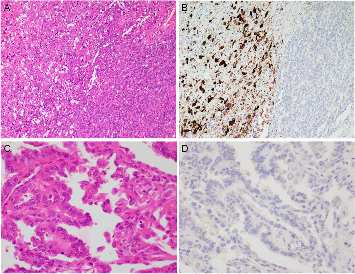
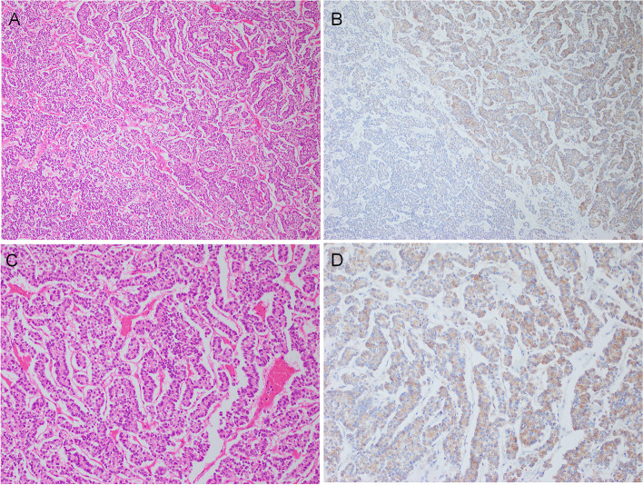
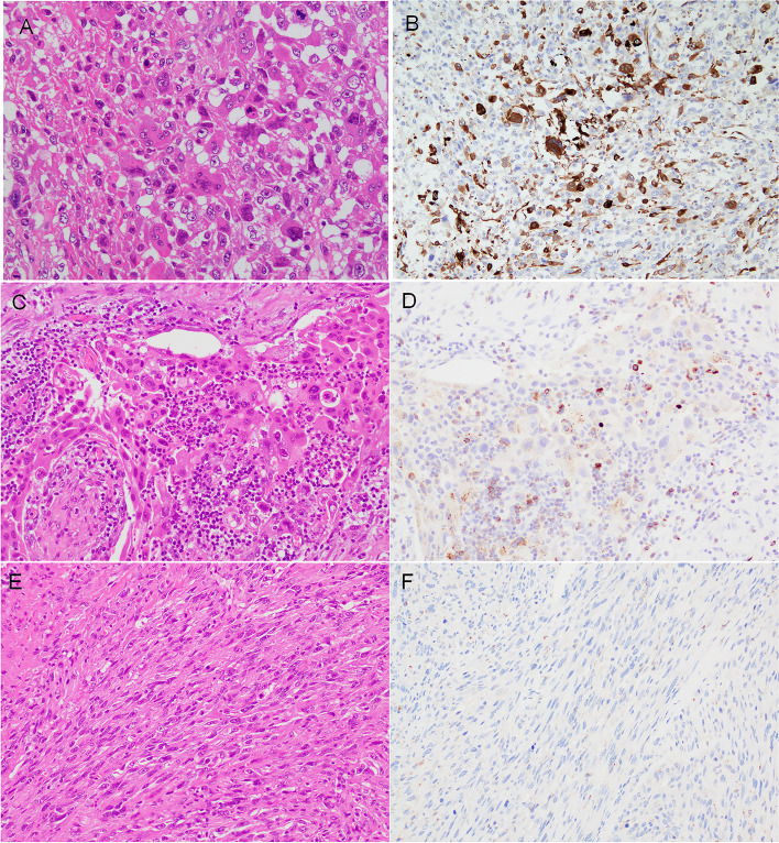
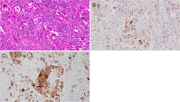
Our IHC results showed that TIGIT expression was detected in cancer cells of MTC and high-grade TC: poorly differentiated thyroid carcinoma (PDTC) and ATC. Neoplastic cells were positive for TIGIT in four of five MTCs (80.0%), 17 of 31 ATCs (54.8%) and in 3 of 12 PDTCs (25.0%). TIGIT was not detected in any adenomatous goiters, thyroid benign tumors, or differentiated thyroid carcinoma (DTCs). Intriguingly, ATC cells showing pleomorphic/giant cell features were positive for TIGIT, while ATC cells with other cell morphologies lacked the immunoreactivity. Intra-tumoral immune cell was inclined to be enriched in TIGI-positive ATC. Although coexisting papillary thyroid carcinoma (PTC) components demonstrated high-grade microscopic features, neither the PTC nor follicular thyroid carcinoma (FTC) components expressed TIGT in any composite ATCs.
TIGIT was immunohistochemically found in MTC with high frequency and partially in high-grade TC. TIGIT expression in cancer cells may be beneficial for a potential utility in MTC and a subset of high-grade TC, especially ATC therapy.
免疫检查点蛋白在滤泡细胞衍生的甲状腺癌和甲状腺髓样癌(MTC)中尚未被充分研究。间变性甲状腺癌(ATC)是最具侵袭性的癌之一。即使采用多模式治疗,ATC 患者的临床结局也不理想。因此,抗肿瘤免疫已被认为对 ATC 具有治疗潜力。
我们使用免疫组织化学(IHC)检查了各种甲状腺病变中的一种新型免疫检查点受体 T 细胞免疫受体免疫球蛋白和酪氨酸基抑制基序域(TIGIT):腺瘤性甲状腺肿、滤泡性腺瘤和甲状腺癌(TC)。
我们的 IHC 结果显示,MTC 和高级 TC(未分化甲状腺癌(PDTC)和 ATC)的癌细胞中检测到 TIGIT 表达。在 5 例 MTC 中的 4 例(80.0%)、31 例 ATC 中的 17 例(54.8%)和 12 例 PDTC 中的 3 例(25.0%)中,肿瘤细胞呈 TIGIT 阳性。在任何腺瘤性甲状腺肿、甲状腺良性肿瘤或分化型甲状腺癌(DTC)中均未检测到 TIGIT。有趣的是,显示多形性/巨细胞特征的 ATC 细胞呈 TIGIT 阳性,而具有其他细胞形态的 ATC 细胞则缺乏免疫反应性。TIGI 阳性的 ATC 中倾向于富集肿瘤内免疫细胞。尽管共存的甲状腺乳头状癌(PTC)成分表现出高级别微观特征,但在任何复合 ATC 中,PTC 或滤泡状甲状腺癌(FTC)成分均不表达 TIGT。
TIGIT 在 MTC 中免疫组化发现频率较高,在部分高级 TC 中也有发现。癌细胞中 TIGIT 的表达可能对 MTC 和高级 TC 亚组(尤其是 ATC)的治疗有益。