Departamento de Neurologia, Faculdade de Ciências Médicas, Universidade de Campinas, Campinas, SP, Brasil.
BRAINN (Brazilian Institute of Neuroscience and Neurotechnology), Campinas, SP, Brasil.
Braz J Med Biol Res. 2022 Aug 15;55:e12036. doi: 10.1590/1414-431X2022e12036. eCollection 2022.
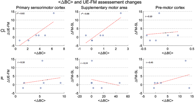
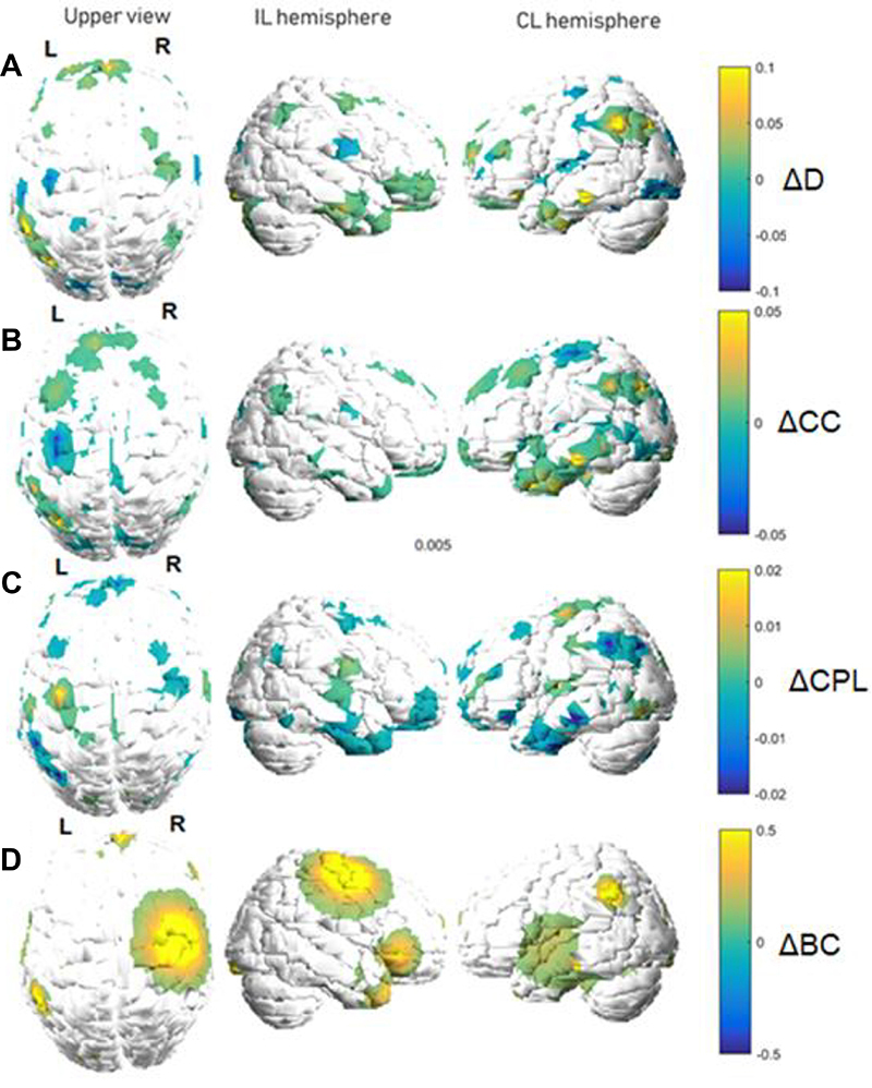
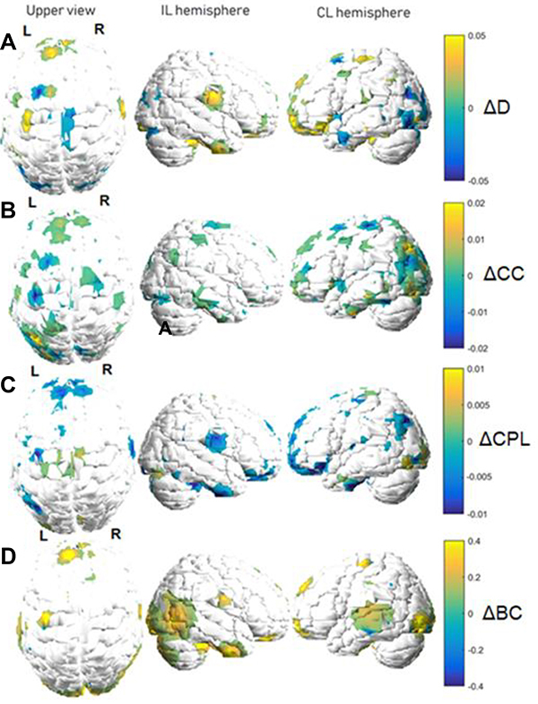
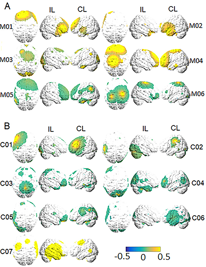
The study of functional reorganization following stroke has been steadily growing supported by advances in neuroimaging techniques, such as functional magnetic resonance imaging (fMRI). Concomitantly, graph theory has been increasingly employed in neuroscience to model the brain's functional connectivity (FC) and to investigate it in a variety of contexts. The aims of this study were: 1) to investigate the reorganization of network topology in the ipsilesional (IL) and contralesional (CL) hemispheres of stroke patients with (motor stroke group) and without (control stroke group) motor impairment, and 2) to predict motor recovery through the relationship between local topological variations of the functional network and increased motor function. We modeled the brain's FC as a graph using fMRI data, and we characterized its interactions with the following graph metrics: degree, clustering coefficient, characteristic path length, and betweenness centrality (BC). For both patient groups, BC yielded the largest variations between the two analyzed time points, especially in the motor stroke group. This group presented significant correlations (P<0.05) between average BC changes and the improvements in upper-extremity Fugl-Meyer (UE-FM) scores at the primary sensorimotor cortex and the supplementary motor area for the CL hemisphere. These regions participate in processes related to the selection, planning, and execution of movement. Generally, higher increases in average BC over these areas were related to larger improvements in UE-FM assessment. Although the sample was small, these results suggest the possibility of using BC as an indication of brain plasticity mechanisms following stroke.
该研究在神经影像学技术(如功能磁共振成像(fMRI))的支持下,对中风后功能重组的研究稳步增长。与此同时,图论在神经科学中被越来越多地用于模拟大脑的功能连接,并在各种情况下对其进行研究。本研究的目的是:1)研究有(运动性中风组)和没有(对照组)运动障碍的中风患者对侧半球(IL)和同侧半球(CL)网络拓扑结构的重组,2)通过功能网络局部拓扑变化与运动功能增强之间的关系预测运动恢复。我们使用 fMRI 数据将大脑的 FC 建模为一个图,并使用以下图度量来描述其相互作用:度、聚类系数、特征路径长度和介数中心性(BC)。对于两个患者组,BC 在两个分析时间点之间产生了最大的变化,特别是在运动性中风组。该组在 CL 半球的初级感觉运动皮层和辅助运动区的平均 BC 变化与上肢 Fugl-Meyer(UE-FM)评分的改善之间存在显著相关性(P<0.05)。这些区域参与与运动选择、计划和执行相关的过程。通常,这些区域的平均 BC 增加幅度越大,UE-FM 评估的改善幅度越大。尽管样本量较小,但这些结果表明,BC 可能作为中风后大脑可塑性机制的一个指标。