Department of Neurosurgery, College of Medicine, Medical University of South Carolina, 96 Jonathan Lucas St., CSB301 MSC606, Charleston, SC, 29425, USA.
Department of Neuroscience, College of Graduate Studies, Medical University of South Carolina, Charleston, SC, USA.
Sci Rep. 2024 Aug 20;14(1):19334. doi: 10.1038/s41598-024-70083-5.
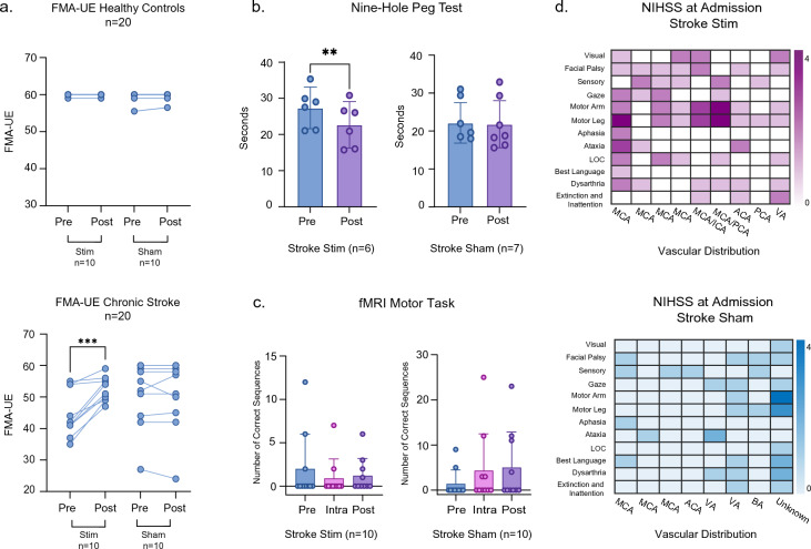
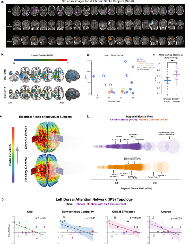
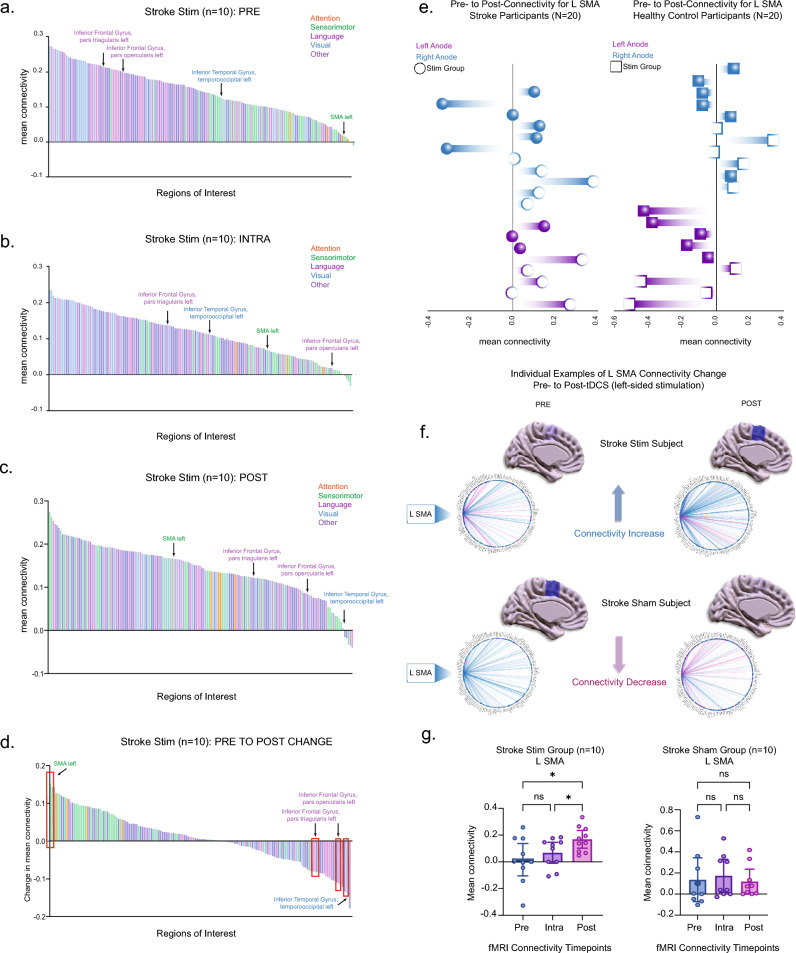
Restoring motor function after stroke necessitates involvement of numerous cognitive systems. However, the impact of damage to motor and cognitive network organization on recovery is not well understood. To discover correlates of successful recovery, we explored imaging characteristics in chronic stroke subjects by combining noninvasive brain stimulation and fMRI. Twenty stroke survivors (6 months or more after stroke) were randomly assigned to a single session of transcranial direct current stimulation (tDCS) or sham during image acquisition. Twenty healthy subjects were included as controls. tDCS was limited to 10 min at 2 mA to serve as a mode of network modulation rather than therapeutic delivery. Fugl-Meyer Assessments (FMA) revealed significant motor improvement in the chronic stroke group receiving active stimulation (p = 0.0005). Motor changes in this group were correlated in a data-driven fashion with imaging features, including functional connectivity (FC), surface-based morphometry, electric field modeling and network topology, focusing on relevant regions of interest. We observed stimulation-related changes in FC in supplementary motor (p = 0.0029), inferior frontal gyrus (p = 0.0058), and temporo-occipital (p = 0.0095) areas, though these were not directly related to motor improvement. The feature most strongly associated with FMA improvement in the chronic stroke cohort was graph topology of the dorsal attention network (DAN), one of the regions surveyed and one with direct connections to each of the areas with FC changes. Chronic stroke subjects with a greater degree of motor improvement had lower signal transmission cost through the DAN (p = 0.029). While the study was limited by a small stroke cohort with moderate severity and variable lesion location, these results nevertheless suggest a top-down role for higher order areas such as attention in helping to orchestrate the stroke recovery process.
中风后恢复运动功能需要涉及众多认知系统。然而,运动和认知网络组织损伤对恢复的影响尚不清楚。为了发现成功恢复的相关因素,我们通过结合无创脑刺激和 fMRI 来探索慢性中风患者的成像特征。20 名中风幸存者(中风后 6 个月或更长时间)在图像采集过程中随机分配到单次经颅直流电刺激(tDCS)或假刺激。20 名健康受试者被纳入对照组。tDCS 限制在 10 分钟内,电流强度为 2 mA,以作为网络调节而不是治疗输送的模式。Fugl-Meyer 评估(FMA)显示接受主动刺激的慢性中风组运动功能显著改善(p=0.0005)。该组的运动变化以数据驱动的方式与成像特征相关,包括功能连接(FC)、基于表面的形态测量学、电场建模和网络拓扑结构,重点关注相关的感兴趣区域。我们观察到补充运动区(p=0.0029)、下额前回(p=0.0058)和颞枕区(p=0.0095)的 FC 发生了与刺激相关的变化,但这些变化与运动改善没有直接关系。与慢性中风队列中 FMA 改善最相关的特征是背侧注意网络(DAN)的拓扑结构,这是调查的区域之一,与具有 FC 变化的区域都有直接连接。DAN 的信号传输成本较低的慢性中风患者运动改善程度更大(p=0.029)。虽然该研究受到中风队列规模较小、中风严重程度中等和病变位置变化的限制,但这些结果表明,像注意这样的高级区域在帮助协调中风恢复过程中起着自上而下的作用。