Department of Ophthalmology, University Hospital Bonn, Bonn, Germany.
University of Bonn, Life & Medical Sciences Institute, Bonn, Germany.
Transl Vis Sci Technol. 2022 Aug 1;11(8):19. doi: 10.1167/tvst.11.8.19.
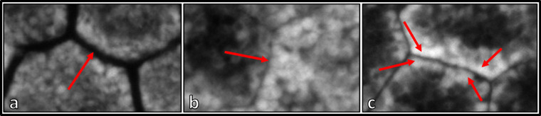
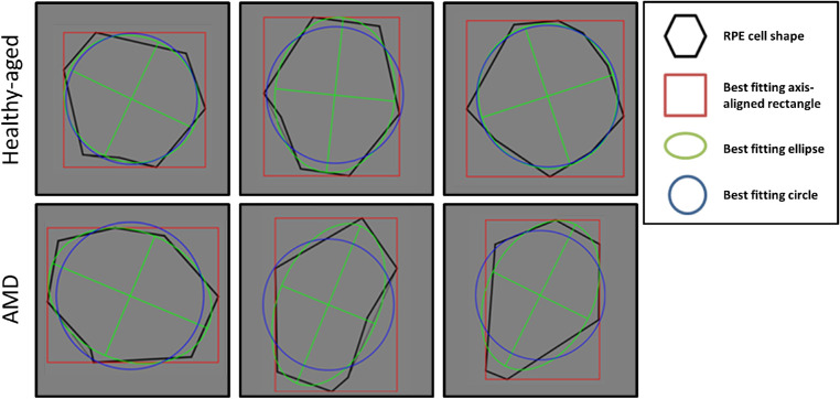
Phenotype alterations of the retinal pigment epithelium (RPE) are a main characteristic of age-related macular degeneration (AMD). Individual RPE cell shape descriptors may help to delineate healthy from AMD-affected cells in early disease stages.
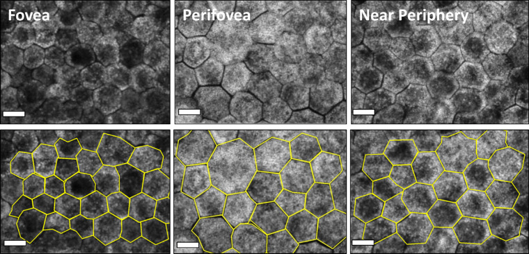
Twenty-two human RPE flatmounts (7 eyes with AMD [early, 3; geographic atrophy, 1; neovascular, 3); 15 unaffected eyes [8 aged ≤51 years; 7 aged >80 years)] were imaged at the fovea, perifovea, and near periphery (predefined sample locations) using a laser-scanning confocal fluorescence microscope. RPE cell boundaries were manually marked with computer assistance. For each cell, 11 shape descriptors were calculated and correlated with donor age, cell autofluorescence (AF) intensity, and retinal location. Statistical analysis was performed using an ensemble classifier based on logistic regression.
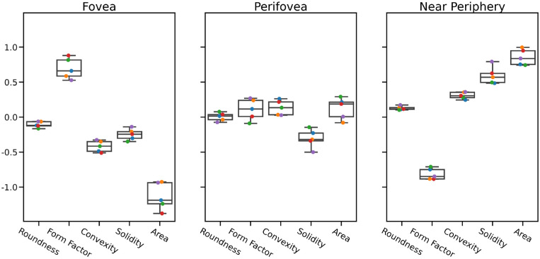
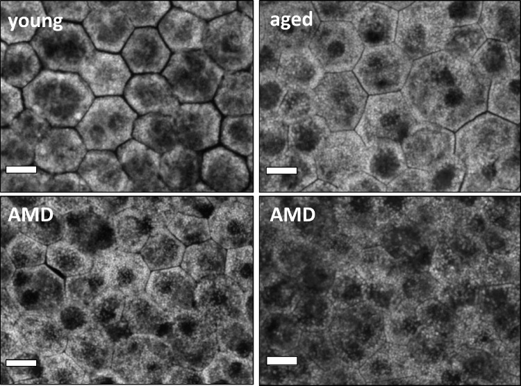
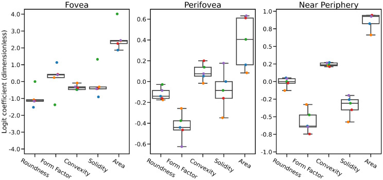
In AMD, RPE was altered at all locations (most pronounced at the fovea), with area, solidity, and form factor being the most discriminatory descriptors. In the unaffected macula, aging had no significant effect on cell shape factors; however, with increasing distance to the fovea, area, solidity, and convexity increased while form factor decreased. Reduced AF in AMD was significantly associated with decreased roundness and solidity.
AMD results in an altered RPE with enlarged and deformed cells that could precede clinically visible lesions and thus serve as early biomarkers for AMD onset. Our data may also help guide the interpretation of RPE morphology in in vivo studies utilizing high-resolution single-cell imaging.
Our histologic RPE cell shape data have the ability to identify robust biomarkers for the early detection of AMD-affected cells, which also could serve as a basis for automated segmentation of RPE sheets.
视网膜色素上皮 (RPE) 的表型改变是年龄相关性黄斑变性 (AMD) 的主要特征。个体 RPE 细胞形状描述符可帮助在疾病早期阶段区分健康细胞和 AMD 相关细胞。
使用激光扫描共聚焦荧光显微镜对 22 个人类 RPE 平面标本(7 只眼睛患有 AMD [早期,3 只;地理萎缩,1 只;新生血管性,3 只];15 只未受影响的眼睛[8 只年龄 ≤51 岁;7 只年龄 >80 岁])的中心凹、中心凹旁和近周边(预定义样本位置)进行成像。使用计算机辅助手动标记 RPE 细胞边界。为每个细胞计算 11 个形状描述符,并与供体年龄、细胞自发荧光 (AF) 强度和视网膜位置相关联。使用基于逻辑回归的集成分类器进行统计分析。
在 AMD 中,RPE 在所有位置都发生了改变(在中心凹处最明显),面积、密实度和形状因子是最具鉴别力的描述符。在未受影响的黄斑区,随着与中心凹距离的增加,细胞面积、密实度和凸度增加,而形状因子减小。AMD 中 AF 的减少与圆度和密实度的降低显著相关。
AMD 导致 RPE 发生改变,细胞增大且变形,这可能早于临床上可见的病变,因此可作为 AMD 发病的早期生物标志物。我们的数据还可能有助于指导利用高分辨率单细胞成像进行的 RPE 形态学的体内研究的解释。
医麦客