Department of Internal Medicine (Nephrology) & Einthoven Laboratory of Vascular and Regenerative Medicine, Leiden University Medical Center, Leiden, the Netherlands.
The Novo Nordisk Foundation Center for Stem Cell Medicine (reNEW), Leiden University Medical Center, Leiden, the Netherlands.
Nat Metab. 2022 Sep;4(9):1109-1118. doi: 10.1038/s42255-022-00615-8. Epub 2022 Aug 25.
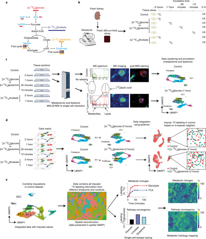
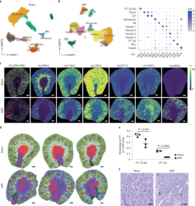
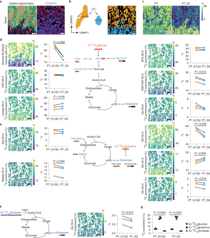
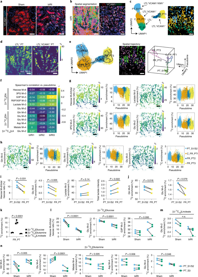
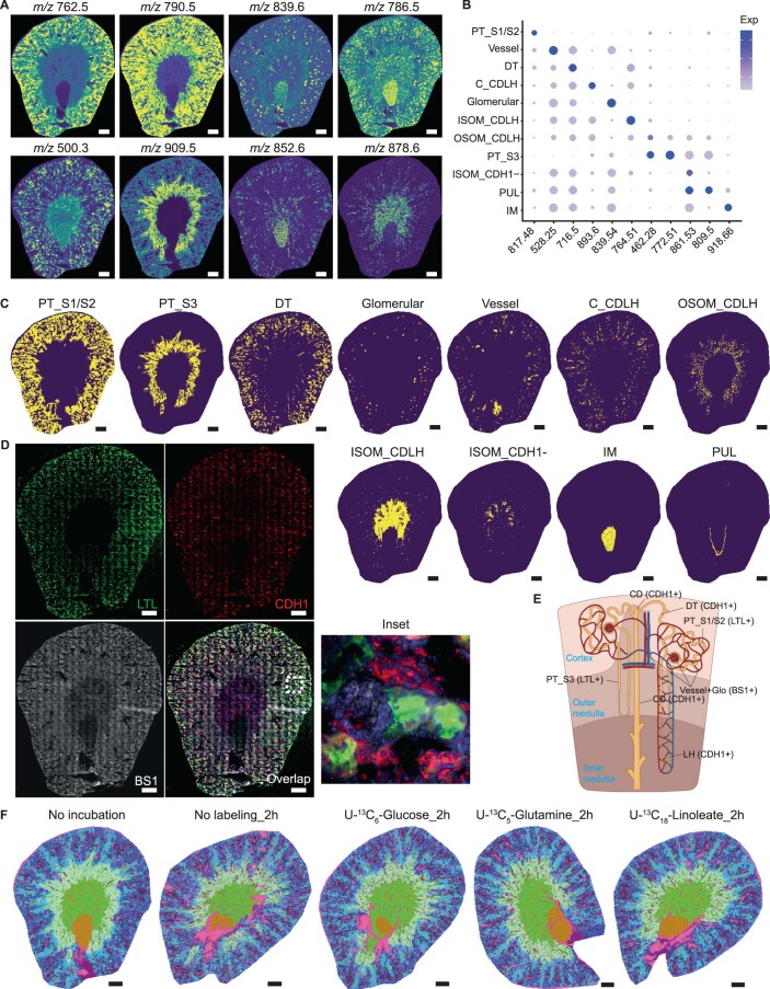
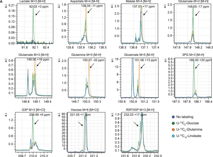
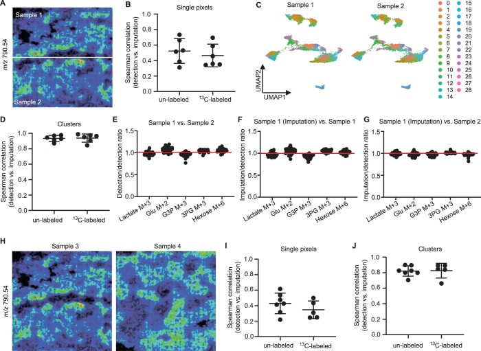
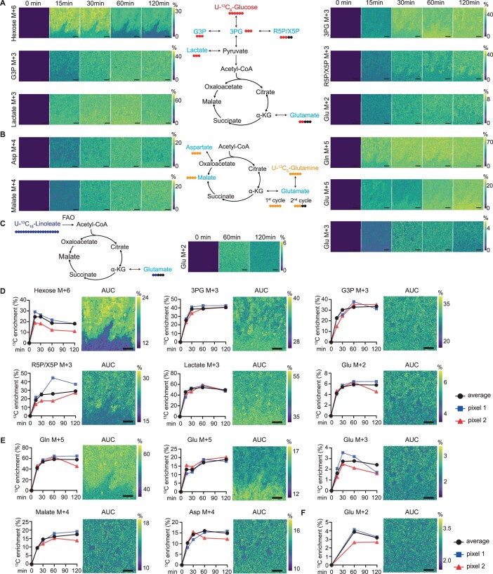
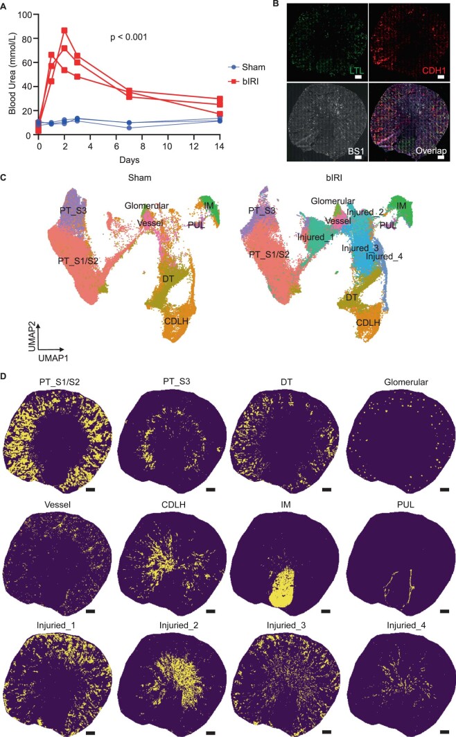
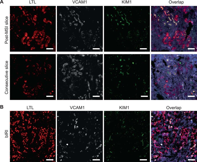
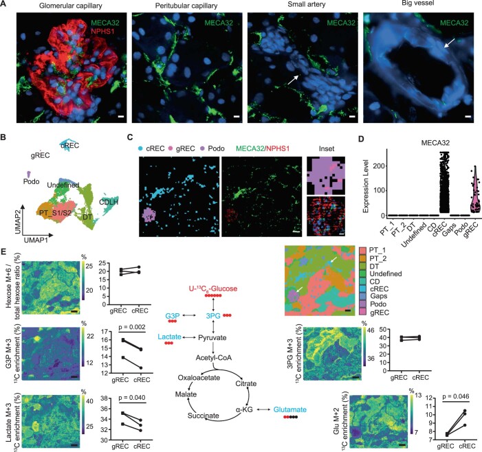
A common drawback of metabolic analyses of complex biological samples is the inability to consider cell-to-cell heterogeneity in the context of an organ or tissue. To overcome this limitation, we present an advanced high-spatial-resolution metabolomics approach using matrix-assisted laser desorption/ionization mass spectrometry imaging (MALDI-MSI) combined with isotope tracing. This method allows mapping of cell-type-specific dynamic changes in central carbon metabolism in the context of a complex heterogeneous tissue architecture, such as the kidney. Combined with multiplexed immunofluorescence staining, this method can detect metabolic changes and nutrient partitioning in targeted cell types, as demonstrated in a bilateral renal ischemia-reperfusion injury (bIRI) experimental model. Our approach enables us to identify region-specific metabolic perturbations associated with the lesion and throughout recovery, including unexpected metabolic anomalies in cells with an apparently normal phenotype in the recovery phase. These findings may be relevant to an understanding of the homeostatic capacity of the kidney microenvironment. In sum, this method allows us to achieve resolution at the single-cell level in situ and hence to interpret cell-type-specific metabolic dynamics in the context of structure and metabolism of neighboring cells.
代谢分析复杂生物样本的一个常见缺点是无法在器官或组织的背景下考虑细胞间的异质性。为了克服这一限制,我们提出了一种先进的高空间分辨率代谢组学方法,使用基质辅助激光解吸/电离质谱成像(MALDI-MSI)结合同位素示踪。该方法允许在复杂的异质组织架构(如肾脏)背景下对中央碳代谢的细胞类型特异性动态变化进行映射。结合多重免疫荧光染色,该方法可以检测靶向细胞类型中的代谢变化和营养分配,如在双侧肾缺血再灌注损伤(bIRI)实验模型中所示。我们的方法使我们能够识别与病变相关的特定区域的代谢扰动,以及整个恢复过程中的代谢扰动,包括在恢复阶段具有明显正常表型的细胞中出现意外的代谢异常。这些发现可能与理解肾脏微环境的稳态能力有关。总之,该方法允许我们在原位达到单细胞水平的分辨率,并因此可以解释细胞类型特异性代谢动力学在邻近细胞的结构和代谢背景下的情况。