Department of Biomedical and Molecular Sciences, Queen's University, Kingston, ON, Canada.
Department of Obstetrics and Gynecology, University of North Carolina, Chapel Hill, NC, Canada.
Front Immunol. 2022 Aug 9;13:961599. doi: 10.3389/fimmu.2022.961599. eCollection 2022.
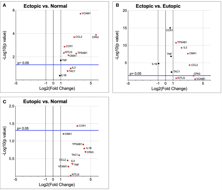
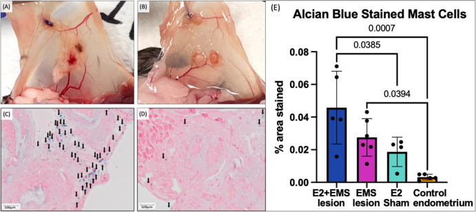
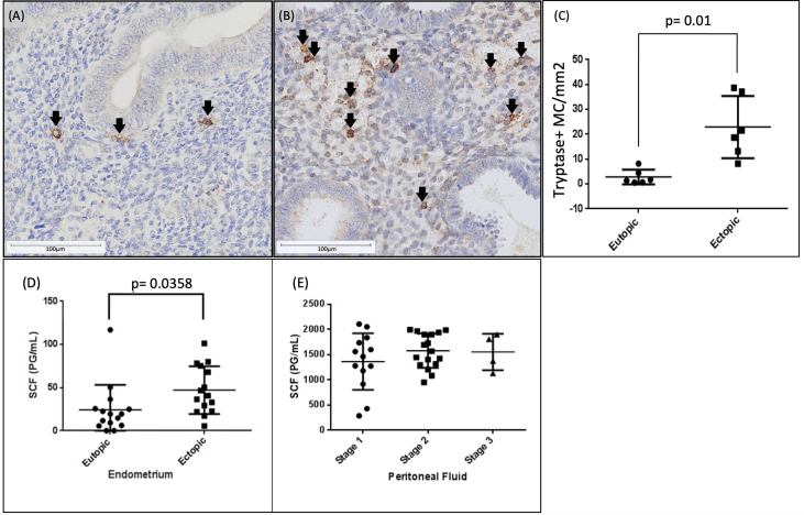
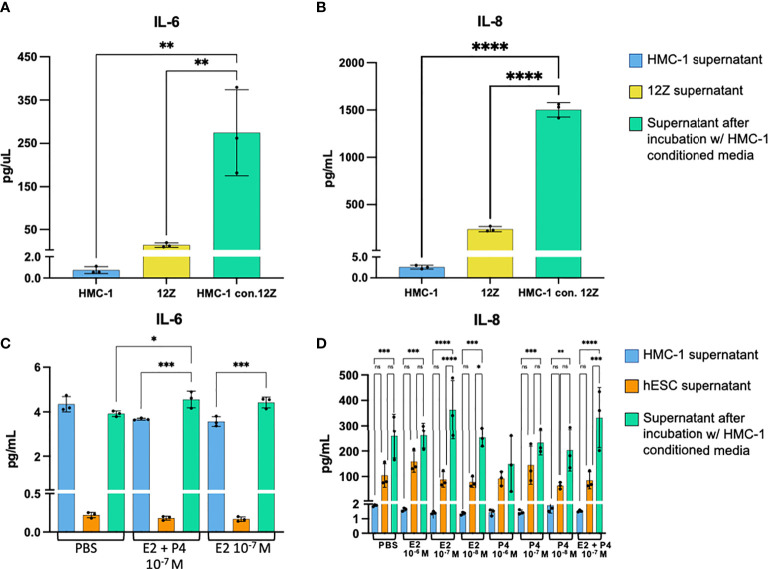
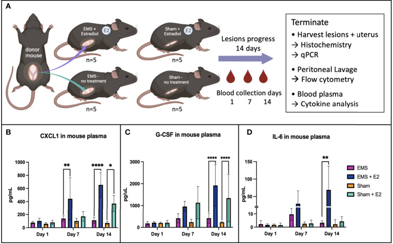
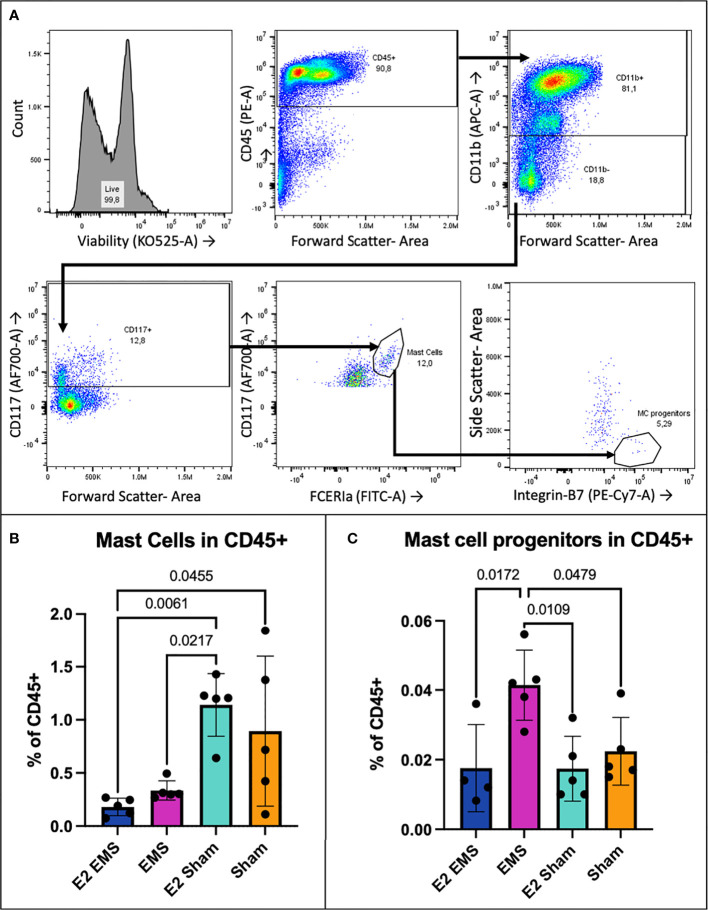
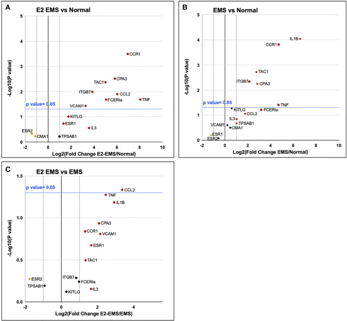
Endometriosis is an estrogen dependent, chronic inflammatory disease characterized by the growth of endometrial lining outside of the uterus. Mast cells have emerged as key players in regulating not only allergic responses but also other mechanisms such as angiogenesis, fibrosis, and pain. The influence of estrogen on mast cell function has also been recognized as a potential factor driving disease pathophysiology in number of allergic and chronic inflammatory conditions. However, precise information is lacking on the cross talk between endocrine and immune factors within the endometriotic lesions and whether that contributes to the involvement of mast cells with disease pathophysiology. In this study, we observed a significant increase in mast cell numbers within endometriotic lesions compared to matched eutopic endometrium from the same patients. Compared to eutopic endometrium, endometriotic lesions had significantly higher levels of stem cell factor (SCF), a potent growth factor critical for mast cell expansion, differentiation, and survival for tissue resident mast cells. Targeted mRNA Q-PCR array revealed that the endometriotic lesions harbour microenvironment (upregulation of CPA3, VCAM1, CCL2, CMA1, CCR1, and KITLG) that is conducive to mast cells recruitment and subsequent differentiation. To examine cross-talk of mast cells within the endometriotic lesion microenvironment, endometriotic epithelial cells (12Z) and endometrial stromal cells (hESC) incubated with mast cell-conditioned media showed significantly increased production of pro-inflammatory and chemokinetic cytokines. To further understand the impact of estrogen on mast cells in endometriosis, we induced endometriosis in C57BL/6 mice. Mature mast cells were significantly higher in peritoneal fluid of estrogen-treated mice compared to untreated mice within the sham operated groups. Mouse endometriotic lesion tissue revealed several genes (qRT-PCR) relevant in mast cell biology significantly upregulated in the estrogen treated, endometriosis-induced group compared to control endometrium. The endometriotic lesions from estrogen treated mice also had significantly higher density of Alcian blue stained mast cells compared to untreated lesions or control endometrium. Collectively, these findings suggest that endometriotic lesions provide a microenvironment necessary for recruitment and differentiation of mast cells. In turn, mast cells potentially release pro-inflammatory mediators that contribute to chronic pelvic pain and endometriosis disease progression.
子宫内膜异位症是一种雌激素依赖性的慢性炎症性疾病,其特征是子宫内膜在子宫外生长。肥大细胞已成为调节不仅过敏反应,而且其他机制,如血管生成、纤维化和疼痛的关键因素。雌激素对肥大细胞功能的影响也被认为是许多过敏性和慢性炎症性疾病中驱动疾病病理生理学的潜在因素。然而,关于子宫内膜异位症病变内内分泌和免疫因素之间的串扰以及这是否导致肥大细胞参与疾病病理生理学的信息仍然缺乏。在这项研究中,我们观察到与同一患者的匹配在位子宫内膜相比,子宫内膜异位症病变中的肥大细胞数量显著增加。与在位子宫内膜相比,子宫内膜异位症病变中干细胞因子 (SCF) 的水平明显更高,SCF 是一种对肥大细胞增殖、分化和存活至关重要的有效生长因子,用于组织驻留肥大细胞。靶向 mRNA Q-PCR 阵列显示,子宫内膜异位症病变具有有利于肥大细胞募集和随后分化的微环境(上调 CPA3、VCAM1、CCL2、CMA1、CCR1 和 KITLG)。为了研究肥大细胞在子宫内膜异位症病变微环境中的串扰,用肥大细胞条件培养基孵育子宫内膜异位症上皮细胞 (12Z) 和子宫内膜基质细胞 (hESC) 后,显示促炎和趋化细胞因子的产生明显增加。为了进一步了解雌激素对子宫内膜异位症中肥大细胞的影响,我们在 C57BL/6 小鼠中诱导子宫内膜异位症。与未治疗的假手术组相比,用雌激素处理的小鼠腹腔液中的成熟肥大细胞明显更高。与对照子宫内膜相比,经雌激素处理、诱导子宫内膜异位症的小鼠的子宫内膜异位症病变组织中与肥大细胞生物学相关的几个基因(qRT-PCR)显著上调。用雌激素处理的小鼠的子宫内膜异位症病变中的阿尔辛蓝染色肥大细胞密度也明显高于未处理的病变或对照子宫内膜。总之,这些发现表明子宫内膜异位症病变提供了募集和分化肥大细胞所需的微环境。反过来,肥大细胞可能释放促炎介质,导致慢性盆腔疼痛和子宫内膜异位症疾病进展。