State Key Laboratory of Molecular Biology, Shanghai Institute of Biochemistry and Cell Biology, Center for Excellence in Molecular Cell Science, Chinese Academy of Sciences, Shanghai 200031, China.
University of Chinese Academy of Sciences, Beijing 100049, China.
Sci Adv. 2022 Aug 26;8(34):eabq4722. doi: 10.1126/sciadv.abq4722.
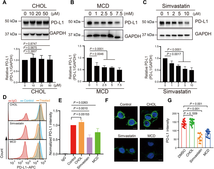
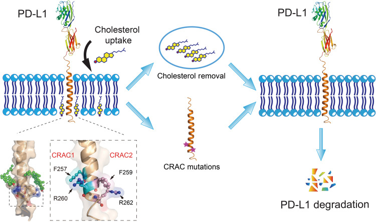
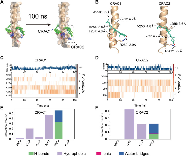
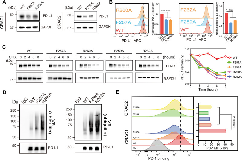
Cholesterol, an essential molecule for cell structure, function, and viability, plays crucial roles in the development, progression, and survival of cancer cells. Earlier studies have shown that cholesterol-lowering drugs can inhibit the high expression of programmed-death ligand 1 (PD-L1) that contributes to immunoevasion in cancer cells. However, the regulatory mechanism of cell surface PD-L1 abundance by cholesterol is still controversial. Here, using nuclear magnetic resonance and biochemical techniques, we demonstrated that cholesterol can directly bind to the transmembrane domain of PD-L1 through two cholesterol-recognition amino acid consensus (CRAC) motifs, forming a sandwich-like architecture and stabilizing PD-L1 to prevent downstream degradation. Mutations at key binding residues prohibit PD-L1-cholesterol interactions, decreasing the cellular abundance of PD-L1. Our results reveal a unique regulatory mechanism that controls the stability of PD-L1 in cancer cells, providing an alternative method to overcome PD-L1-mediated immunoevasion in cancers.
胆固醇是细胞结构、功能和存活所必需的分子,在癌细胞的发展、进展和存活中发挥着关键作用。早期研究表明,降低胆固醇的药物可以抑制程序性死亡配体 1(PD-L1)的高表达,从而有助于癌细胞的免疫逃逸。然而,胆固醇对细胞表面 PD-L1 丰度的调节机制仍存在争议。在这里,我们使用核磁共振和生化技术证明,胆固醇可以通过两个胆固醇识别氨基酸共识(CRAC)基序直接与 PD-L1 的跨膜结构域结合,形成三明治样结构,稳定 PD-L1 以防止下游降解。关键结合残基的突变会阻止 PD-L1-胆固醇相互作用,从而降低 PD-L1 的细胞丰度。我们的结果揭示了一种控制癌细胞中 PD-L1 稳定性的独特调节机制,为克服癌症中 PD-L1 介导的免疫逃逸提供了一种替代方法。