• 文献检索
  • 文档翻译
  • 深度研究
  • 学术资讯
  • Suppr Zotero 插件Zotero 插件
  • 邀请有礼
  • 套餐&价格
  • 历史记录
应用&插件
Suppr Zotero 插件Zotero 插件浏览器插件Mac 客户端Windows 客户端微信小程序
定价
高级版会员购买积分包购买API积分包
服务
文献检索文档翻译深度研究API 文档MCP 服务
关于我们
关于 Suppr公司介绍联系我们用户协议隐私条款
关注我们

Suppr 超能文献

核心技术专利:CN118964589B侵权必究
粤ICP备2023148730 号-1Suppr @ 2026

文献检索

告别复杂PubMed语法,用中文像聊天一样搜索,搜遍4000万医学文献。AI智能推荐,让科研检索更轻松。

立即免费搜索

文件翻译

保留排版,准确专业,支持PDF/Word/PPT等文件格式,支持 12+语言互译。

免费翻译文档

深度研究

AI帮你快速写综述,25分钟生成高质量综述,智能提取关键信息,辅助科研写作。

立即免费体验

活样本的多尺度荧光成像。

Multiscale fluorescence imaging of living samples.

机构信息

Laboratory of High-Resolution Optical Imaging, National Institute of Biomedical Imaging and Bioengineering, National Institutes of Health, Bethesda, MD, 20892, USA.

Janelia Research Campus, Howard Hughes Medical Institute, Ashburn, VA, 20147, USA.

出版信息

Histochem Cell Biol. 2022 Oct;158(4):301-323. doi: 10.1007/s00418-022-02147-4. Epub 2022 Aug 29.

DOI:10.1007/s00418-022-02147-4
PMID:36036808
原文链接:https://pmc.ncbi.nlm.nih.gov/articles/PMC9512758/
Abstract

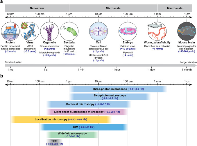
Fluorescence microscopy is a highly effective tool for interrogating biological structure and function, particularly when imaging across multiple spatiotemporal scales. Here we survey recent innovations and applications in the relatively understudied area of multiscale fluorescence imaging of living samples. We discuss fundamental challenges in live multiscale imaging and describe successful examples that highlight the power of this approach. We attempt to synthesize general strategies from these test cases, aiming to help accelerate progress in this exciting area.

摘要

荧光显微镜是研究生物结构和功能的一种非常有效的工具,特别是在跨多个时空尺度成像时。在这里,我们调查了相对研究较少的活样本多尺度荧光成像领域的最新创新和应用。我们讨论了活多尺度成像中的基本挑战,并描述了成功的例子,突出了这种方法的优势。我们试图从这些案例中综合出一般策略,旨在帮助加速这一令人兴奋的领域的进展。

https://cdn.ncbi.nlm.nih.gov/pmc/blobs/b528/9512758/64995f1c316d/418_2022_2147_Fig9_HTML.jpg
https://cdn.ncbi.nlm.nih.gov/pmc/blobs/b528/9512758/a3531b362fce/418_2022_2147_Fig1_HTML.jpg
https://cdn.ncbi.nlm.nih.gov/pmc/blobs/b528/9512758/82e6bc9d4547/418_2022_2147_Fig2_HTML.jpg
https://cdn.ncbi.nlm.nih.gov/pmc/blobs/b528/9512758/1e432b7c874a/418_2022_2147_Fig3_HTML.jpg
https://cdn.ncbi.nlm.nih.gov/pmc/blobs/b528/9512758/a60cf0c2df24/418_2022_2147_Fig4_HTML.jpg
https://cdn.ncbi.nlm.nih.gov/pmc/blobs/b528/9512758/af2d1ffeb4bb/418_2022_2147_Fig5_HTML.jpg
https://cdn.ncbi.nlm.nih.gov/pmc/blobs/b528/9512758/66c762ffa89f/418_2022_2147_Fig6_HTML.jpg
https://cdn.ncbi.nlm.nih.gov/pmc/blobs/b528/9512758/66f697d263bb/418_2022_2147_Fig7_HTML.jpg
https://cdn.ncbi.nlm.nih.gov/pmc/blobs/b528/9512758/77c27ab90bb5/418_2022_2147_Fig8_HTML.jpg
https://cdn.ncbi.nlm.nih.gov/pmc/blobs/b528/9512758/64995f1c316d/418_2022_2147_Fig9_HTML.jpg

相似文献

1
Multiscale fluorescence imaging of living samples.活样本的多尺度荧光成像。
Histochem Cell Biol. 2022 Oct;158(4):301-323. doi: 10.1007/s00418-022-02147-4. Epub 2022 Aug 29.
2
Light sheet microscopy and live imaging of plants.植物的光片显微镜技术与活体成像
J Microsc. 2016 Aug;263(2):158-64. doi: 10.1111/jmi.12393. Epub 2016 Mar 28.
3
STED super-resolved microscopy.受激发射损耗超分辨显微镜。
Nat Methods. 2018 Mar;15(3):173-182. doi: 10.1038/nmeth.4593. Epub 2018 Jan 29.
4
Quantum dots in bioanalysis: a review of applications across various platforms for fluorescence spectroscopy and imaging.量子点在生物分析中的应用:综述荧光光谱和成像在各种平台上的应用。
Appl Spectrosc. 2013 Mar;67(3):215-52. doi: 10.1366/12-06948.
5
Diffraction-unlimited fluorescence microscopy of living biological samples using pcSOFI.使用相位相关超分辨光学涨落成像技术对活的生物样本进行无衍射荧光显微镜成像。
Curr Protoc Chem Biol. 2015 Mar 2;7(1):27-41. doi: 10.1002/9780470559277.ch140025.
6
Localizing protein-protein interactions in living cells using fluorescence lifetime imaging microscopy.利用荧光寿命成像显微镜在活细胞中定位蛋白质-蛋白质相互作用。
Methods Mol Biol. 2015;1251:83-107. doi: 10.1007/978-1-4939-2080-8_6.
7
Super-resolution fluorescence imaging of directly labelled DNA: from microscopy standards to living cells.直接标记 DNA 的超分辨率荧光成像:从显微镜标准到活细胞。
J Microsc. 2013 Jul;251(1):1-4. doi: 10.1111/jmi.12054. Epub 2013 May 23.
8
Microscopy in Infectious Disease Research-Imaging Across Scales.感染病研究中的显微镜检查法——跨尺度成像。
J Mol Biol. 2018 Aug 17;430(17):2612-2625. doi: 10.1016/j.jmb.2018.06.018. Epub 2018 Jun 24.
9
Follow-up review: recent progress in the development of super-resolution optical microscopy.随访综述:超分辨率光学显微镜发展的最新进展
Microscopy (Oxf). 2016 Aug;65(4):275-81. doi: 10.1093/jmicro/dfw022. Epub 2016 Jul 6.
10
Single-molecule fluorescence imaging in living cells.活细胞中的单分子荧光成像。
Annu Rev Phys Chem. 2013;64:459-80. doi: 10.1146/annurev-physchem-040412-110127. Epub 2013 Jan 16.

引用本文的文献

1
A Multimodal Adaptive Optical Microscope For Imaging from Molecules to Organisms.一种用于从分子到生物体成像的多模态自适应光学显微镜。
bioRxiv. 2025 Jun 3:2025.06.02.657494. doi: 10.1101/2025.06.02.657494.
2
Quantifying UV-induced photodamage for longitudinal live-cell imaging applications of deep-UV microscopy.用于深紫外显微镜纵向活细胞成像应用的紫外线诱导光损伤定量分析。
Biomed Opt Express. 2024 Dec 19;16(1):208-221. doi: 10.1364/BOE.544778. eCollection 2025 Jan 1.
3
Lineage Tracing by Light-Sheet Microscopy and Computational Reconstruction.

本文引用的文献

1
Capturing the start point of the virus-cell interaction with high-speed 3D single-virus tracking.高速 3D 单病毒追踪捕获病毒-细胞相互作用的起始点。
Nat Methods. 2022 Dec;19(12):1642-1652. doi: 10.1038/s41592-022-01672-3. Epub 2022 Nov 10.
2
Incorporating the image formation process into deep learning improves network performance.将图像形成过程纳入深度学习可以提高网络性能。
Nat Methods. 2022 Nov;19(11):1427-1437. doi: 10.1038/s41592-022-01652-7. Epub 2022 Oct 31.
3
Event-driven acquisition for content-enriched microscopy.
通过光片显微镜和计算重建进行谱系追踪
Methods Mol Biol. 2025;2886:153-176. doi: 10.1007/978-1-0716-4310-5_8.
4
A 'double-edged' role for type-5 metabotropic glutamate receptors in pain disclosed by light-sensitive drugs.光敏感药物揭示了 5 型代谢型谷氨酸受体在疼痛中的“双刃剑”作用。
Elife. 2024 Aug 22;13:e94931. doi: 10.7554/eLife.94931.
5
Label-free functional analysis of root-associated microbes with dynamic quantitative oblique back-illumination microscopy.利用动态定量斜向背照显微镜对根相关微生物进行无标记功能分析。
Sci Rep. 2024 Mar 9;14(1):5812. doi: 10.1038/s41598-024-56443-1.
6
Live-cell imaging powered by computation.基于计算的活细胞成像。
Nat Rev Mol Cell Biol. 2024 Jun;25(6):443-463. doi: 10.1038/s41580-024-00702-6. Epub 2024 Feb 20.
7
A "double-edged" role for type-5 metabotropic glutamate receptors in pain disclosed by light-sensitive drugs.光敏药物揭示了5型代谢型谷氨酸受体在疼痛中的“双刃剑”作用。
bioRxiv. 2024 Jan 3:2024.01.02.573945. doi: 10.1101/2024.01.02.573945.
8
Soft X-ray Microscopy in Cell Biology: Current Status, Contributions and Prospects.细胞生物学中的软X射线显微镜:现状、贡献与展望
Acta Naturae. 2023 Oct-Dec;15(4):32-43. doi: 10.32607/actanaturae.26551.
9
Imagining the future of optical microscopy: everything, everywhere, all at once.想象光学显微镜的未来:一切、无处不在、一切都在瞬间。
Commun Biol. 2023 Oct 28;6(1):1096. doi: 10.1038/s42003-023-05468-9.
10
In focus in HCB.在六氯苯研究中成为焦点。
Histochem Cell Biol. 2022 Oct;158(4):297-299. doi: 10.1007/s00418-022-02149-2.
基于事件驱动的获取技术在富含内容的显微镜中的应用
Nat Methods. 2022 Oct;19(10):1262-1267. doi: 10.1038/s41592-022-01589-x. Epub 2022 Sep 8.
4
Event-triggered STED imaging.事件触发的 STED 成像。
Nat Methods. 2022 Oct;19(10):1268-1275. doi: 10.1038/s41592-022-01588-y. Epub 2022 Sep 8.
5
Stereotyped behavioral maturation and rhythmic quiescence in embryos.胚胎刻板的行为成熟和节律静止。
Elife. 2022 Aug 5;11:e76836. doi: 10.7554/eLife.76836.
6
Single-molecule localization microscopy.单分子定位显微镜技术
Nat Rev Methods Primers. 2021;1. doi: 10.1038/s43586-021-00038-x. Epub 2021 Jun 3.
7
Combining multiple fluorescence imaging techniques in biology: when one microscope is not enough.在生物学中结合多种荧光成像技术:当一台显微镜不够用时。
Mol Biol Cell. 2022 May 15;33(6):tp1. doi: 10.1091/mbc.E21-10-0506.
8
Caveat fluorophore: an insiders' guide to small-molecule fluorescent labels.警示荧光团:小分子荧光标记物的内部人士指南。
Nat Methods. 2022 Feb;19(2):149-158. doi: 10.1038/s41592-021-01338-6. Epub 2021 Dec 23.
9
Multiview confocal super-resolution microscopy.多视场共聚焦超分辨显微镜。
Nature. 2021 Dec;600(7888):279-284. doi: 10.1038/s41586-021-04110-0. Epub 2021 Nov 26.
10
Imaging intact human organs with local resolution of cellular structures using hierarchical phase-contrast tomography.利用分层相衬断层摄影术,以局部分辨率对完整的人体器官进行成像。
Nat Methods. 2021 Dec;18(12):1532-1541. doi: 10.1038/s41592-021-01317-x. Epub 2021 Nov 4.