Media Lab, MIT, Cambridge, MA, USA.
MIT Center for Neurobiological Engineering, MIT, Cambridge, MA, USA.
Nat Biomed Eng. 2022 Sep;6(9):1057-1073. doi: 10.1038/s41551-022-00912-3. Epub 2022 Aug 29.
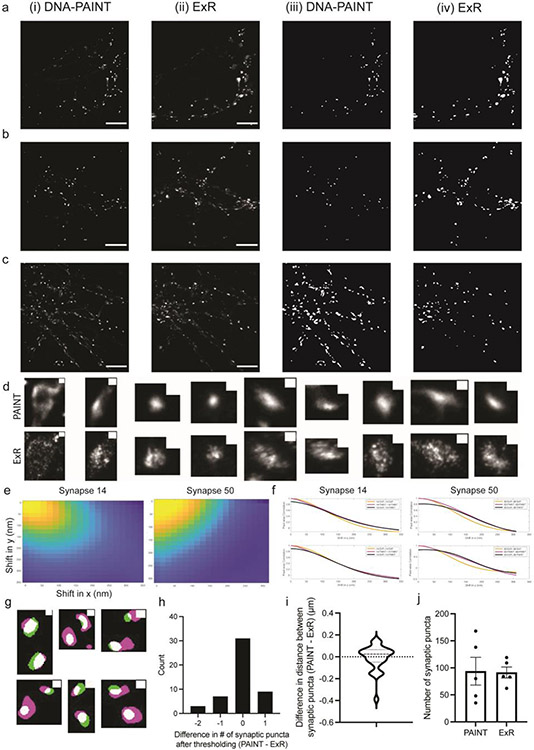
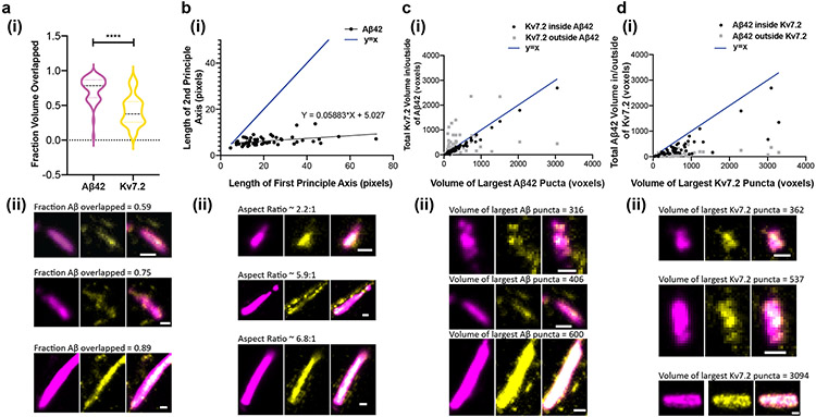
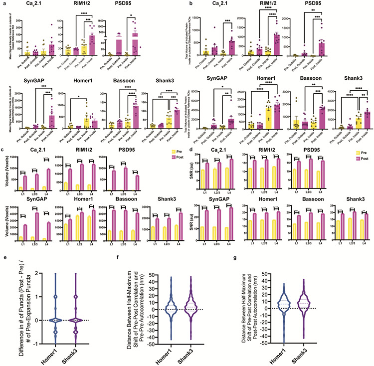
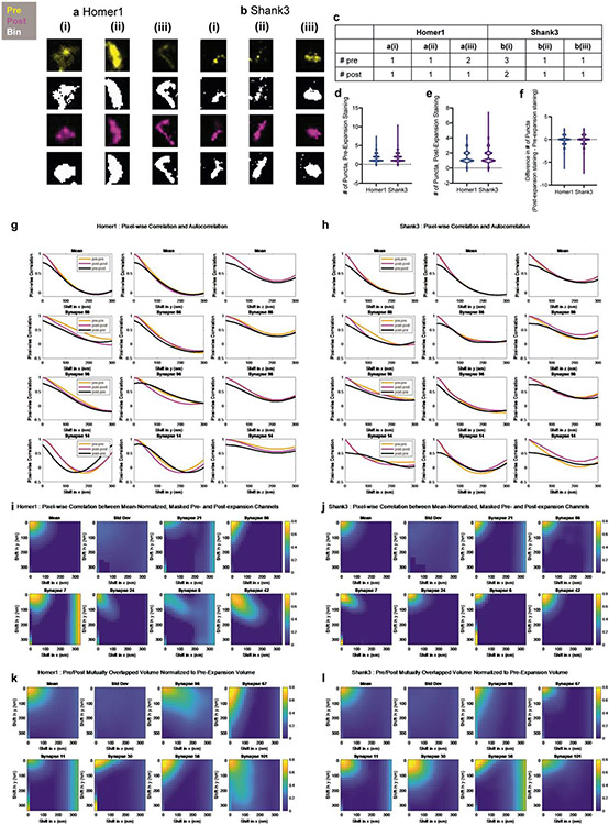
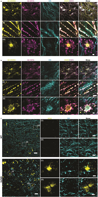
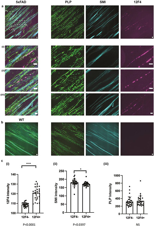
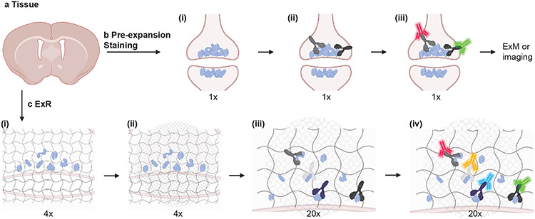
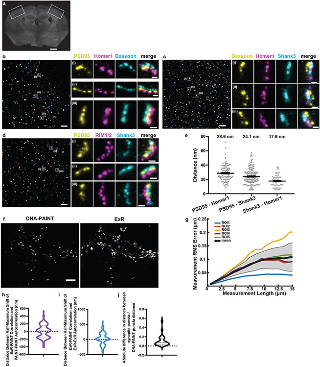
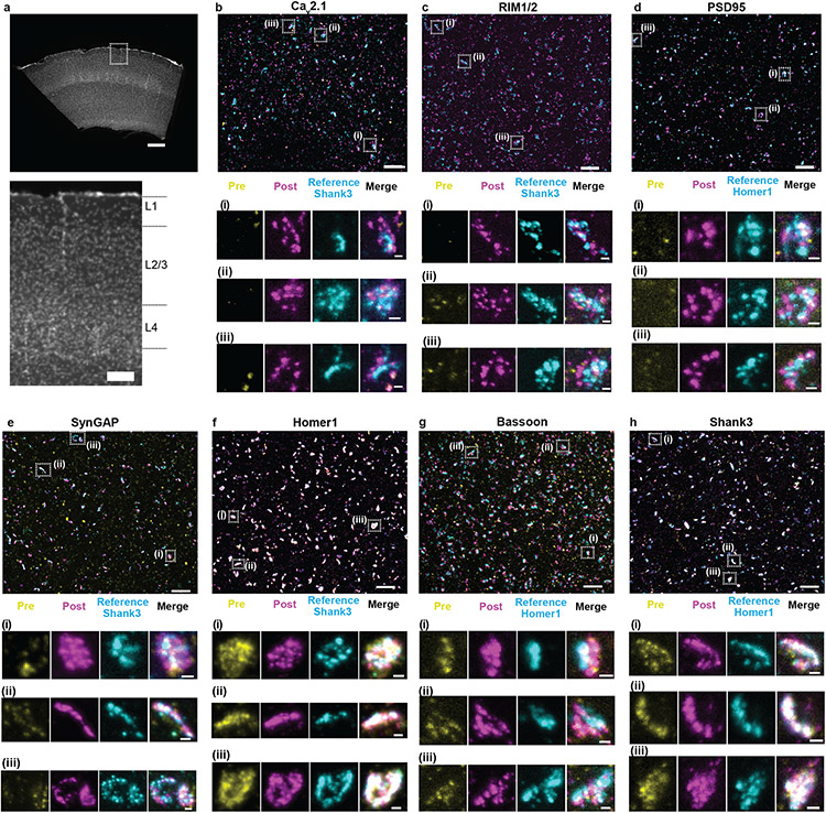
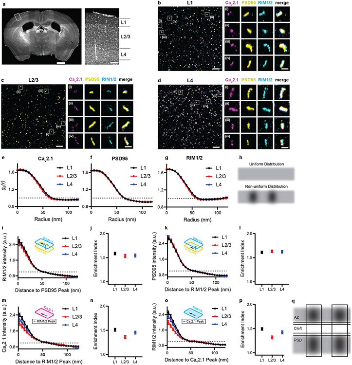
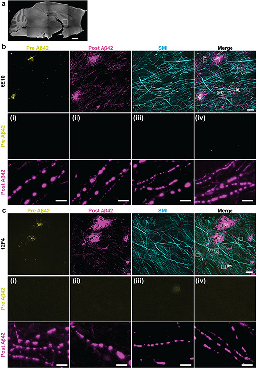
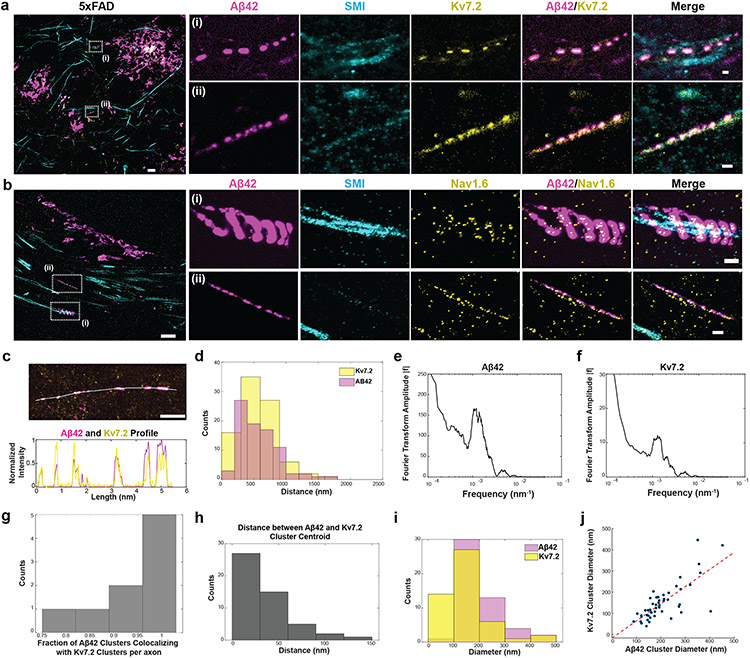
Many crowded biomolecular structures in cells and tissues are inaccessible to labelling antibodies. To understand how proteins within these structures are arranged with nanoscale precision therefore requires that these structures be decrowded before labelling. Here we show that an iterative variant of expansion microscopy (the permeation of cells and tissues by a swellable hydrogel followed by isotropic hydrogel expansion, to allow for enhanced imaging resolution with ordinary microscopes) enables the imaging of nanostructures in expanded yet otherwise intact tissues at a resolution of about 20 nm. The method, which we named 'expansion revealing' and validated with DNA-probe-based super-resolution microscopy, involves gel-anchoring reagents and the embedding, expansion and re-embedding of the sample in homogeneous swellable hydrogels. Expansion revealing enabled us to use confocal microscopy to image the alignment of pre-synaptic calcium channels with post-synaptic scaffolding proteins in intact brain circuits, and to uncover periodic amyloid nanoclusters containing ion-channel proteins in brain tissue from a mouse model of Alzheimer's disease. Expansion revealing will enable the further discovery of previously unseen nanostructures within cells and tissues.
许多细胞和组织中拥挤的生物分子结构无法被标记抗体标记。因此,为了了解这些结构内的蛋白质如何以纳米级精度排列,需要在标记之前对这些结构进行去拥挤处理。在这里,我们展示了一种迭代的扩展显微镜方法(细胞和组织通过可溶胀水凝胶进行渗透,然后进行各向同性水凝胶扩展,以允许使用普通显微镜实现更高的成像分辨率),可以在扩展但保持完整的组织中以约 20nm 的分辨率成像纳米结构。该方法我们称为“扩展揭示”,并通过 DNA 探针超分辨率显微镜进行了验证,涉及凝胶锚定试剂以及在均匀可溶胀水凝胶中对样品的嵌入、扩展和重新嵌入。扩展揭示使我们能够使用共聚焦显微镜来观察完整脑回路中突触前钙离子通道与突触后支架蛋白的排列,并在阿尔茨海默病小鼠模型的脑组织中揭示含有离子通道蛋白的周期性淀粉样纳米簇。扩展揭示将能够进一步发现细胞和组织内以前看不见的纳米结构。